
Compromiso torácico de la sarcoidosis en tomografía computarizada: claves clínicas y radiológicas
DOI:
https://doi.org/10.53903/01212095.5Palabras clave:
Tomografía computarizada multidetector, Radiografía torácica, Sarcoidosis, Sarcoidosis pulmonarResumen
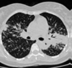
La sarcoidosis es una enfermedad crónica y multisistémica de etiología poco clara. La presentación es variable, de acuerdo con la procedencia geográfica del paciente, pero predomina en personas afrodescendientes y escandinavas. Las anormalidades torácicas son muy frecuentes en los pacientes con sarcoidosis; la afectación más común es ganglionar y la menos común es la del corazón. Las manifestaciones radiológicas más frecuentes en el compromiso torácico por sarcoidosis son las adenomegalias hiliares y mediastinales, así como nódulos pulmonares de distribución perilinfática.
Descargas
Referencias bibliográficas
Lee GM, Pope K, Meek L, Chung JH, Hobbs SB, Walker CM. Sarcoidosis: A diagnosis of exclusion. AJR. 2020;214:1-9. https://doi.org/10.2214/AJR.19.21436
Prabhakar HB, Rabinowitz CB, Gibbons FK, O'Donnell WJ, Shepard JO, Aquino SL. Imaging features of sarcoidosis on MDCT, FDG PET, and PET/CT. AJR. 2008;190:S1-S6. https://doi.org/10.2214/AJR.07.7001
Semionov A, Kosiuk J, Ajlan AM, Discepola F. Thoracic diseases with musculoske- letal manifestations and vice versa: A review. AJR. 2018;211:1000-9. https://doi.org/10.2214/AJR.18.19803
Ganeshan D, Menias CO, Lubner MG, Pickhardt PJ, Sandrasegaran K, Bhalla S. Sarcoidosis from head to toe: what the radiologist needs to know. Radiographics. 2018;38(4):1180-200. https://doi.org/10.1148/rg.2018170157
Reddy GP, Ahuja J. Thoracic sarcoidosis: Imaging patterns. Sem Roentgenol. 2019;54(1):59-65. https://doi.org/10.1053/j.ro.2018.12.008
Akaike G, Itani M, Shah H, et al. PET/CT in the diagnosis and workup of sarcoidosis: Focus on atypical manifestations. RadioGraphics. 2018;38:1536-49. https://doi.org/10.1148/rg.2018180053
Ortega IH, González LL. Update thoracic sarcoidosis. Radiología. 2011;53(5):434- 48. https://doi.org/10.1016/j.rx.2011.03.010
Silva M, Nunes H, Valeyre D, Sverzellati N. Imaging of sarcoidosis. Clinic Rev Allerg Immunol. 2015;49:45-53. https://doi.org/10.1007/s12016-015-8478-7
Nunes H, Brillet PY, Valeyre D, Brauner MW, Wells AU. Imaging in sarcoidosis. Semin Respir Crit Care Med. 2007;28(1):102-20. https://doi.org/10.1055/s-2007-970336
Nunes H, Uzunhan Y, Gille T, Lamberto C, Valeyre D, Brillet PY. Imaging of sarcoidosis of the airways and lung parenchyma and correlation with lung function. Eur Respir J. 2012;40:750-65. https://doi.org/10.1183/09031936.00025212
Spagnolo P, Sverzellati N, Wells AU, Hansell DM. Imaging aspects of the diagnosis of sarcoidosis. Eur Radiol. 2014;24:807-16. https://doi.org/10.1007/s00330-013-3088-3
Keijsers RG, Veltkamp M, Grutters JC. Chest imaging. Clin Chest Med. 2015;36:603-19. https://doi.org/10.1016/j.ccm.2015.08.004
Hawtin KE, Roddie ME, Mauri FA, Copley SJ. Pulmonary sarcoidosis: the 'Great Pretender'. Clin Radiol. 2010;65:642-50. https://doi.org/10.1016/j.crad.2010.03.004
Criado E, Sánchez M, Ramírez J, Arguis P, de Caralt TM, Perea RJ, et al. Pulmonary sarcoidosis: typical and atypical manifestations at high-resolution CT with pathologic correlation. RadioGraphics. 2010;30:1567-86. https://doi.org/10.1148/rg.306105512
Conant EF, Glickstein MF, Mahar P, Miller WT. Pulmonary sarcoidosis in the older patient: conventional radiographic features. Radiology. 1988;169(2):315-9. https://doi.org/10.1148/radiology.169.2.3174979
Nunes H, Humbert M, Capron F, Brauner M, Sitbon O, Battesti JP, et al. Pulmonary hypertension associated with sarcoidosis: mechanisms, haemodynamics and prognosis. Thorax. 2006;61:68-74. https://doi.org/10.1136/thx.2005.042838
Walsh SL, Wells AU, Sverzellati N, Keir GJ, Calandriello L, AntoniouKM, et al. An integrated clinicoradiological staging system for pulmonary sarcoidosis: a case-cohort study. Lancet Respir Med. 2014;2(2):123-30. https://doi.org/10.1016/S2213-2600(13)70276-5
Rosen Y, Moon S, Huang CT, Gourin A, Lyons HA. Granulomatous pulmonary angiitis in sarcoidosis. Arch Pathol Lab Med. 1977;101:170-4.
Takemura T, Matsui Y, Saiki S, Mikami R. Pulmonary vascular involvement in sarcoidosis: a report of 40 autopsy cases. Hum Pathol. 1992;23:1216-23. https://doi.org/10.1016/0046-8177(92)90288-E
Hoffstein V, Ranganathan N, Mullen JB. Sarcoidosis simulating pulmonary veno-occlusive disease. Am Rev Respir Dis. 1986;134:809-11. doi: 10.1164/arrd.1986.134.4.809.
Jones RM, Dawson A, Jenkins GH, Nicholson AG, Hansell DM, Harrison NK. Sarcoidosis-related pulmonary veno-occlusive disease presenting with recurrent haemoptysis. Eur Respir J. 2009;34:517-20. https://doi.org/10.1183/09031936.00044609
Corte TJ, Wells AU, Nicholson AG, Hansell DM, Wort SJ. Pulmonary hypertension in sarcoidosis: A review. Respirology. 2011;16:69-77. https://doi.org/10.1111/j.1440-1843.2010.01872.x
Simonneau G, Gatzoulis MA, Adatia I, Celermajer D, Denton C, Ghofrani A, et al. Updated clinical classification of pulmonary hypertension. J Am Coll Cardiol. 2013;62(25 suppl):D34-D41. https://doi.org/10.1016/j.jacc.2013.10.029
Aluja-Jaramillo F, Gutiérrez FR, Díaz-Telli F, Yevenes-Aravena S, Javidan-Nejad C, Bhalla S. Approach to pulmonary hypertension: From CT to clinical diagnosis. Radiographics. 2018;38(2):357-73. https://doi.org/10.1148/rg.2018170046
Koo HJ, Chae EJ, Kim JE, Kim EY, Oh SY, Hwang HJ. Presence of macronodules in thoracic sarcoidosis: prevalence and computed tomographic findings. Clin Radiol. 2015;70(8):815-21. https://doi.org/10.1016/j.crad.2015.03.012
Koyama T, Ueda H, Togashi K, Umeoka S, Kataoka M, Nagai S. Radiologic manifestations of sarcoidosis in various organs. RadioGraphics. 2004;24(1):87-104. https://doi.org/10.1148/rg.241035076
Müller NL, Kullnig P, Miller RR. The CT findings of pulmonary sarcoidosis: analysis of 25 patients. AJR Am J Roentgenol. 1989;152(6):1179-82. https://doi.org/10.2214/ajr.152.6.1179
Hamper UM, Fishman EK, Khouri NF, Johns CJ, Wang KP, Siegelman SS. Typical and atypical CT manifestations of pulmonary sarcoidosis. J Comput Assist Tomogr. 1986;10(6):928-36. https://doi.org/10.1097/00004728-198611000-00006
Crystal RG, Roberts WC, Hunninghake GW, Gadek JE, Fulmer JD, Line BR. Pulmonary sarcoidosis: a disease characterized and perpetuated by activated lung T-lymphocytes. Ann Intern Med. 1981;94:73-94. https://doi.org/10.7326/0003-4819-94-1-73
Bonham CA, Strek ME, Patterson KC. From granuloma to fibrosis: sarcoidosis as- sociated pulmonary fibrosis. Curr Opin Pulm Med. 2016;22(5):484-91. https://doi.org/10.1097/MCP.0000000000000301
Patterson KC, Strek ME. Pulmonary fibrosis in sarcoidosis. Clinical features and outcomes. Annals ATS. 2013;10(4):362-70. https://doi.org/10.1513/AnnalsATS.201303-069FR
Gerke AK. Morbidity and mortality in sarcoidosis. Curr Opin Pulm Med. 2014;20(5):472-8. https://doi.org/10.1097/MCP.0000000000000080
Viskum K, Vestbo J. Vital prognosis in intrathoracic sarcoidosis with special reference to pulmonary function and radiological stage. Eur Respir J. 1993;6:349-53.
Shlobin OA, Nathan SD. Management of end-stage sarcoidosis: pulmonary hypertension and lung transplantation. Eur Respir J. 2012;39(6):1520-33. https://doi.org/10.1183/09031936.00175511
Abehsera M, Valerye D, Greiner P, Jaillet H, Battesti JP, Brauner MW. Sarcoidosis with pulmonary fibrosis: CT patterns and correlation with pulmonary function. AJR Am J Roentgenol. 2000;174(6):1751-7. https://doi.org/10.2214/ajr.174.6.1741751
Grenier P, Valeyre D, Cluzel P, Brauner MW, Lenoir S, Chastang C. Chronic diffuse interstitial lung disease: diagnostic value of chest radiography and high-resolution CT. Radiology. 1991;179:123-32. https://doi.org/10.1148/radiology.179.1.2006262
Grenier P, Chevret S, Beigelman C, Brauner MW, Chastang C, Valeyre D. Chronic diffuse infiltrative lung disease: determination of the diagnostic value of clinical data, chest radiography, and CT with Bayesian analysis. Radiology. 1994;191: 383-90. https://doi.org/10.1148/radiology.191.2.8153310
Balan A, Hoey ETD, Sheerin F, Lakkaraju A, Chowdhury FU. Multi-technique imaging of sarcoidosis. Clin Radiol. 2010;65:750-60. https://doi.org/10.1016/j.crad.2010.03.014
Nakatsu M, Hatabu H, Morikawa K, Uematsu H, Ohno Y, Nishimura K, et al. Large coalescent parenchymal nodules in pulmonary sarcoidosis: "sarcoid galaxy" sign. AJR Am J Roentgenol. 2002;178(6):1389-93. https://doi.org/10.2214/ajr.178.6.1781389
Gotway MB, Tchao NK, Leung JW, Hanks DK, Thomas AN. Sarcoidosis presenting as an enlarging solitary pulmonary nodule. J Thorac Imaging. 2001;16(2):117-22. https://doi.org/10.1097/00005382-200104000-00010
Herráez Ortega I, Alonso Orcajo N, López González L. The "sarcoid cluster sign": a new sign in high resolution chest CT [in Spanish]. Radiología. 2009;51(5):495-9. https://doi.org/10.1016/j.rx.2009.05.003
Polychronopoulos VS, Prakash UBS. Airway involvement in sarcoidosis. Chest. 2009;136:1371-80. https://doi.org/10.1378/chest.08-2569
Aluja-Jaramillo F, Gutiérrez FR, Rossi S, Bhalla S. Nonneoplastic tracheal abnormalities on CT. Contempor Diagn Radiol. 2020:43;13:1-7. https://doi.org/10.1097/01
Park HJ, Jung JI, Chung MH, Song SW, Kim HL, Baik JH, et al. Typical and Atypical Manifestations of Intrathoracic Sarcoidosis. Korean J Radiol.2009;10:623-631. https://doi.org/10.3348/kjr.2009.10.6.623
Soskel NT, Sharma OP. Pleural involvement in sarcoidosis: case presentation and detailed review of the literature. Semin Respir Med. 1992;13(6):492-514. https://doi.org/10.1055/s-2007-1006299
Lynch JP III, Kazerooni EA, Gay SE. Pulmonary sarcoidosis. Clin Chest Med. 1997;18(4):755-85. https://doi.org/10.1016/S0272-5231(05)70417-2
Jarman PR, Whyte MK, Sabroe I, Hughes JM. Sarcoidosis presenting with chylothorax. Thorax. 1995;50(12):1324-5. https://doi.org/10.1136/thx.50.12.1324
Blankstein R, Waller AH. Evaluation of known or suspected cardiac sarcoidosis. Circ Cardiovasc Imaging. 2016;9:e000867. https://doi.org/10.1161/CIRCIMAGING.113.000867
Birnie DH, Sauer WH, Bogun F, Cooper JM, Culver DA, Duvernoy S, et al. HRS expert consensus statement on the diagnosis and management of arrhythmias as- sociated with cardiac sarcoidosis. Heart Rhythm. 2014;11:1305-23. https://doi.org/10.1016/j.hrthm.2014.03.043
Sobic-Saranovic D, Artiko V, Obradovic V. FDG PET imaging in sarcoidosis. Semin Nucl Med. 2013;43(6):404-11. https://doi.org/10.1053/j.semnuclmed.2013.06.007
Teirstein AS, Machac J, Almeida O, Lu P, Padilla ML, Iannuzzi MC. Results of 188 whole-body fluorodeoxyglucose positron emission tomography scans in 137 patients with sarcoidosis. Chest. 2007;132:1949-53. https://doi.org/10.1378/chest.07-1178
Krüger S, Buck AK, Mottaghy FM, Pauls S, Schelzig H, Hombach V, et al. Use of integrated FDG-PET/CT in sarcoidosis. Clin Imaging. 2008;32(4):269-73. https://doi.org/10.1016/j.clinimag.2007.11.005
Love C, Tomas MB, Tronco GG, Palestro CJ. FDG PET of infection and inflammation. RadioGraphics. 2005;25:1357-68. https://doi.org/10.1148/rg.255045122
Braun JJ, Kessler R, Constantinesco A, Imperiale A. 18F-FDG PET/CT in sarcoidosis management: review and report of 20 cases. Eur J Nucl Med Mol Imaging. 2008;35:1537-43. https://doi.org/10.1007/s00259-008-0770-9
Descargas
Publicado
Cómo citar
Número
Sección
Licencia
Derechos de autor 2020 Revista Colombiana de Radiología

Esta obra está bajo una licencia internacional Creative Commons Atribución-NoComercial-CompartirIgual 4.0.
La Revista Colombiana de Radiología es de acceso abierto y todos sus artículos se encuentran libre y completamente disponibles en línea para todo público sin costo alguno.
Los derechos patrimoniales de autor de los textos y de las imágenes del artículo como han sido transferidos pertenecen a la Asociación Colombiana de Radiología (ACR). Por tanto para su reproducción es necesario solicitar permisos y se debe hacer referencia al artículo de la Revista Colombiana de Radiología en las presentaciones o artículos nuevos donde se incluyan.


