
Ultrasonido de los nervios periféricos de la extremidad inferior
DOI:
https://doi.org/10.53903/01212095.143Palabras clave:
Ultrasonido, Fibras nerviosas, Extremidad inferiorResumen
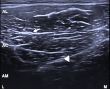
Los recientes avances en los equipos de ultrasonido y de sus transductores han permitido la mayor competitividad del método, posicionándose como la primera opción sobre otras modalidades de imagen en la valoración de las enfermedades de los tendones y nervios, gracias al incremento en la resolución para visualizar y explorar los tendones y los nervios periféricos. El ultrasonido (US) se consideraba un complemento de la resonancia magnética (RM); sin embargo, con los equipos modernos, se ha convertido en la mejor modalidad diagnóstica para la revisión de los nervios periféricos, aceptada cada día más, debido a la rapidez, disponibilidad y la característica dinámica del método ecográfico. La desventaja principal es que depende del operador y que la experiencia de quien lo realiza es fundamental para su adquisición e interpretación. Este trabajo muestra una breve revisión de la técnica y de los marcadores anatómicos en la valoración de los nervios periféricos más comunes de la extremidad inferior.
Descargas
Referencias bibliográficas
Tagliafico A, Bignotti B, Martinoli C. Update on ultrasound-guided interventional procedures on peripheral nerves semin musculoskelet. Radiol. 2016;20:453-60.
Martinoli C, Bianchi S, Dahmane M, Pugliese F. et al. Ultrasound of tendons and nerves. Eur Radiol. 2002;12:44-55.
Tagliafico AS. Peripheral nerve imaging: Not only cross-sectional área. World J Radiol.2016;8:726-28.
Martinoli C, Bianchi S, Derchi LE. Tendon and nerve sonography. Radiol Clin North Am.1999;37:691-711.
Tagliafico A, Martinoli C. Reliability of sonographic measurements of upper extremity nerves. J Ultrasound Med. 2013;32:457-62.
Vlassakov KV, Sala Bach X. Ultrasound of the peripheral nerves. Nerves Nerve Inj. 2015;1:227-50.
Tagliafico AS, Tagliafico G. Fascicular ratio: a new parameter to evaluate peripheral nerve pathology on magnetic resonance imaging: a feasibility study on a 3T MRI system. Medicine (Baltimore). 2014;93:e68.
Bedewi MA, Abodonya A, Kotb M, Kamal S, et al. Estimation of ultrasound reference values for the lower limb peripheral nerves in adults: A cross-sectional study. Medicine. 2018;97(12):e0179.
Suk JI, Walker FO, Cartwright MS. Ultrasound of peripheral nerves. Curr Neurol Neurosci Rep. 2013;13:328.
Tagliafico AS, González RP, Rossi F, Bignotti B, Martinoli C. Peripheral nerves: Not only cross-sectional area in the era of radiomics. Semin Musculoskelet Radiol. 2020;24:175-80.
Tagliafico A, Tagliafico G, Martinoli C. Nerve density: a new parameter to evaluate peripheral nerve pathology on ultrasound. Preliminary study. Ultrasound Med Biol 2010; 36:1588-93.
Tagliafico A, Bignotti B. New parameters for evaluating peripheral nerve disorders on sonography and magnetic resonance imaging. J Ultrasound Med. 2015:34:1523.
Tagliafico A, Bignotto B, Rossi F, Sconfienza L, et al. Ultrasound of the hip joint, Soft Tissues and nerves. Semin in Musculoskeletal Radiol. 2017;21:582-8.
Tagliafico, Martinoli C. Lateral femoral cutaneous nerve. J Ultrasound Med. 2011;30:1341-46.
Jacobson JA. Fundamentals of musculoskeletal ultrasound. En Wrist and hand ultrasound. Elsevier; 2018. pp. 174-77.
Yablon CM, Hammer MR, Morag Y, Brandon CJ, Fessell DP, Jacobson JA. US of the peripheral nerves of the lower extremity: A landmark approach. Radiographics. 2016;36:464-78.
Jacobson JA, Wilson TJ, Yang LJS. Sonography of common peripheral nerve disorders. Ultrasound Med. 2016;35:683-93.
Iagnocco A, Filippucci E, Meenagh G, Delle Sedie A, et al. Ultrasound imaging for the rheumatologist. I. Ultrasonography of the shoulder. Clin Exp Rheumatol. 2006;24:6-11.
Hospodar PP, Ashman ES, Traub JA. Anatomic study of the lateral femoral cutaneous nerve with respect to the ilioinguinal surgical dissection. J Orthop Trauma. 1999;13:17-19.
Tagliafico A, Pérez M, Martinoli C. High-resolution ultrasound of the pudendal nerve. Normal anatomy. Muscle Nerve. 2013;47:403-8.
Tagliafico A, Bignotti B, Cadoni A, Pérez MM, Martinoli C. Anatomical study of the iliohypogastric, ilioinguinal, and genitofemoral nerves using high-resolution ultrasound. Muscle Nerve. 2015;51:42-8.
Kowalska B, Sudoł-Szopińska I. Normal and sonographic anatomy of selected peripheral nerves. Part III: Peripheral nerves of the lower limb. J Ultrason. 2012;12:148-63.
Beltran LS, Bencardino J, Ghazikhanian V, Beltran J. Entrapment neuropathies III: lower limb. Semin Musculoskelet Radiol. 2010;14:501-11.
De Maeseneer M, Madami H, Lenchik L, Brígido MK, et al. Normal anatomy and compression areas of nerves of the foot and ankle: US and MR Imaging with anatomic correlation. Radiographics. 2015;35:1469-82.
Presley JC, Maida E, Pawlina W, Murthy N, et al. Sonographic visualization of the first branch of the lateral plantar nerve (baxter nerve): technique and validation using perineural injections in a cadaveric model. J Ultrasound Med. 2013;32:1643-52.
Alshami AM, Souvlis T, Coppieters MW. A review of plantar heel pain of neural origin: differential diagnosis and management. Man Ther. 2008;13:103-11.
Ucerler H, Ikiz ‘A. The variations of the sensory branches of the superficial peroneal nerve course and its clinical importance. Foot Ankle Int. 2005;26:942-6.
Nwawka OK, Lee S, Miller TT. Sonographicevaluation of superficial peroneal nerve abnormalities. AJR 2018;211:1-8.
Descargas
Publicado
Cómo citar
Número
Sección
Licencia
Derechos de autor 2022 Revista colombiana de radiología

Esta obra está bajo una licencia internacional Creative Commons Atribución-NoComercial-CompartirIgual 4.0.
La Revista Colombiana de Radiología es de acceso abierto y todos sus artículos se encuentran libre y completamente disponibles en línea para todo público sin costo alguno.
Los derechos patrimoniales de autor de los textos y de las imágenes del artículo como han sido transferidos pertenecen a la Asociación Colombiana de Radiología (ACR). Por tanto para su reproducción es necesario solicitar permisos y se debe hacer referencia al artículo de la Revista Colombiana de Radiología en las presentaciones o artículos nuevos donde se incluyan.


