
Presentación leptomeníngea primaria de tumores con restricción en difusión. Presentación de dos casos
DOI:
https://doi.org/10.53903/01212095.199Palabras clave:
Neoplasias del sistema nervioso central, Imagen por resonancia magnética, PediatríaResumen
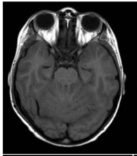
Los tumores leptomeníngeos primarios en pediatría son entidades poco comunes. En su mayoría, se trata de tumores glioneuronales difusos, aunque también se han descrito algunos casos de tumores embrionarios. La complejidad del diagnóstico de esta presentación se debe a las manifestaciones clínicas atípicas y a las dificultades en el diagnóstico diferencial. La secuencia de imagen ponderada por difusión (DWI) es una modalidad de imagen altamente sensible que detecta el movimiento del agua en el espacio extracelular. En neurooncología, su utilidad radica en diferenciar entre tumores de baja densidad celular de aquellos con alta celularidad, particularmente de los que se encuentran compuestos por células “pequeñas, redondas y azules”. En este estudio se describen dos casos de tumores leptomeníngeos primarios diseminados de origen embrionario, sin una masa cerebral primaria, con restricción en la secuencia de DWI (hipercelularidad). Los casos, estudiados en el Hospital Garrahan en los tres últimos años, y la revisión de la literatura indican que el hallazgo imagenológico más frecuente es el engrosamiento y realce nodular leptomeníngeo intracraneal e intraespinal difuso. Sin embargo, no se encontraron informes sobre la utilidad de la secuencia de DWI para el diagnóstico de estas entidades. En el artículo se analizan los enfoques de neuroimagen y la confirmación diagnóstica con el fin de proporcionar oportunidades para un tratamiento efectivo de estas enfermedades en la práctica clínica.
Descargas
Referencias bibliográficas
Louis DN, Perry A, Wesseling P, et al. The 2021 WHO Classification of Tumors of the Central Nervous System: a summary. Neuro Oncol; 2021;23(8):1231-51. https://doi.org/10.1093/neuonc/noab1063
Osborn AG, Louis DN, Poussaint TY, et al. The 2021 World Health Organization Classification of Tumors of the Central Nervous System: What neuroradiologists need to know. AJNR Am J Neuroradiol. 2022;43(7):928-37. https://doi.org/10.3174/ajnr.A7462
Min L, Yuhao D, Wangming Z. Molecular determinants of medulloblastoma metastasis and leptomeningeal dissemination. Mol Cancer Res. 2021;19:743-52. https://doi.org/10.1158/1541-7786.MCR-20-1026
Ríos CI, De Jesus O. Primitive neuroectodermal tumor. StatPearls [Internet]. Treasure Island (FL): StatPearls Publishing; 2022. PMID: 32965836.
Tanaka H, Yamamoto D, Ikeda M, et al. Embryonal brain tumor with unknown primary lesion and massive cerebrospinal fluid dissemination: A case report. J Clin Neurosci. 2018;54:125-8. https://doi.org/10.1016/j.jocn.2018.04.046
Shih R, Koeller K. Embryonal tumors of the central nervous system: From the radiologic pathology archives. RadioGraphics. 2018;38:2:525-41. https://doi.org/10.1148/rg.2018170182
Morgacheva D, Daks A, Smirnova A, et al. Primary leptomeningeal medulloblastoma in a child: Clinical case report and literature review. Front Pediatr. 2022;10:925340. https://doi.org/10.3389/fped.2022.925340
Ferrara M, Bizzozero L, Fiumara E, et al. “Primary” leptomeningeal dissemination of medulloblastoma. Report of an unusual case. J Neurosurg Sci. 1989;33:219-23. PMID: 2795197.
Suman R, Santosh V, Anandh BA. Primary leptomeningeal medulloblastoma. Pediatr Neurosurg. 2007;43:544-5. https://doi.org/10.1159/000108806
Mehta RI, Cutler AR, Lasky III JL, et al. “Primary” leptomeningeal medulloblastoma. Hum Pathol. 2009;40:1661-5. https://doi.org/10.1016/j.humpath.2009.04.024
Ghosh A, Slopis J, Koenig MK. Primary leptomeningeal medulloblastoma: a rare presentation. AAN Ent. 2018;6:105.
Sublett J, Davenport C, Eisenbrock H, et al. Pediatric primary difusse leptomeningeal Primitive Neuroectodermal Tumor: A case report and literature review. Pediatr Neurosurg. 2017;52(2):114-21. https://doi.org/10.1159/000452807
Tomomasa R, Nakata S, Nobusawa S, et al. Primary diffuse leptomeningeal atypical teratoid/rhabdoid tumor diagnosed by cerebrospinal fluid cytology: case report with molecular genetic analysis. Hum Pathol. 2018;77:116-20. https://doi.org/10.1016/j.humpath.2017.12.026
El-Nabbout B, Shbarou R, Glasier CM, et al. Primary diffuse cerebral leptomeningeal atypical teratoid rhabdoid tumor: report of the first case. J Neurooncol. 2010;98:431-4. https://doi.org/10.1007/s11060-009-0094-z
Gauvain KM, Durham BH, MuHugh M, et al. Rapidly progressive primary leptomeningeal Atypical teratoid/rhabdoid tumor: a report of 2 cases. J Child Neurol. 2012;27:1596-601. https://doi.org/10.1177/0883073812436878
Livermore LJ, Dabbous B, Hofer M, et al. Primary diffuse leptomeningeal atypical teratoid/rhabdoid tumour in an adolescent. Clin Neurol Neurosurg. 2013;115:2170-3. https://doi.org/10.1016/j.clineuro.2013.05.036
Surov A, Meyer HJ, Wienke A. Correlation between apparent diffusion coefficient (ADC) and cellularity is different in several tumors: a meta-analysis. Oncotarget. 2017;8(35):59492-9. https://doi.org/10.18632/oncotarget.17752
Rumboldt Z, Camacho DL, Lake D, et al. Apparent diffusion coefficients for differentiation of cerebellar tumors in children. AJNR Am J Neuroradiol. 2006;27(6):1362-9. PMID: 16775298; PMCID: PMC8133915.
Smirniotopoulos JG, Murphy FM, Rushing EJ, et al. Patterns of contrast enhancement in the brain and meninges. RadioGraphics. 2007;27(2):525-51. https://doi.org/10.1148/rg.272065155
Gardiman MP, Fassan M, Orvieto E, et al. Diffuse leptomeningeal glioneuronal tumors: a new entity? Brain Pathol. 2010;20(2):361-6. https://doi.org/10.1111/j.1750-3639.2009.00285
Lakhani DA, Mankad K, Chhabda S, et al. Diffuse leptomeningeal glioneuronal tumor of childhood. AJNR Am J Neuroradiol. 2020;41(11):2155-9. https://doi.org/10.3174/ajnr.A6737
Peer S, Murumkar V, Kulanthaivelu K, et al. Diffuse leptomeningeal glioneuronal tumor with high-grade features masquerading as tubercular meningitis—a case report. Egypt J Radiol Nucl Med. 2021;52:146. https://doi.org/10.1186/s43055-021-00522-0
Amer EM, Youssef AF, Romeih MA, et al. Role of magnetic resonance imaging in characterization of central nervous system lesions in pediatric patients with leukemia and post-treatment complications. Egypt
Descargas
Publicado
Cómo citar
Número
Sección
Licencia
Derechos de autor 2024 Revista colombiana de radiología

Esta obra está bajo una licencia internacional Creative Commons Atribución-NoComercial-CompartirIgual 4.0.
La Revista Colombiana de Radiología es de acceso abierto y todos sus artículos se encuentran libre y completamente disponibles en línea para todo público sin costo alguno.
Los derechos patrimoniales de autor de los textos y de las imágenes del artículo como han sido transferidos pertenecen a la Asociación Colombiana de Radiología (ACR). Por tanto para su reproducción es necesario solicitar permisos y se debe hacer referencia al artículo de la Revista Colombiana de Radiología en las presentaciones o artículos nuevos donde se incluyan.


