
Valoración prequirúrgica de anomalías dentofaciales mediante tomografía axial computarizada: lo que el radiólogo siempre debería informar
DOI:
https://doi.org/10.53903/01212095.230Palabras clave:
Maloclusión, Deformidades dentofaciales, Tomografía computarizada por rayos XResumen
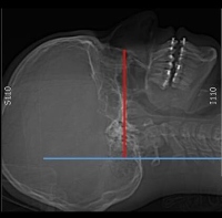
Las anomalías dentofaciales (ADF) son un motivo frecuente de consulta en los servicios de cirugía maxilofacial, puesto que el diagnóstico por imagen es importante para la planificación y el seguimiento quirúrgico. Las ADF se refieren a la desviación significativa de las proporciones normales del complejo maxilomandibular, que afectan negativamente la oclusión dental. El manejo de estas alteraciones se realiza principalmente con cirugía ortognática, que busca el reposicionamiento de los componentes dentales y del esqueleto facial mediante una combinación de procedimientos. Los estudios de tomografía axial computarizada permiten a los cirujanos maxilofaciales establecer el tipo de anomalía, identificar los hallazgos asociados —alteraciones de la respiración—, realizar simulaciones quirúrgicas y definir una planificación quirúrgica precisa. Los grupos de trabajo interdisciplinarios y el conocimiento mínimo del lenguaje utilizado en el manejo de las ADF permiten un mejor abordaje de las modalidades de imagen, así como el reporte de información relevante para el manejo clínico de los pacientes.
Descargas
Referencias bibliográficas
Posnick JC. Principles and practice of the orthognathic surgery. Elsevier Saunders; 2013.
Scarfe WC, Angelopoulos C. Maxillofacial cone beam computed tomography. Springer International Publishing; 2018.
Kim EJ, Ki EJ, Cheon HM, Choi EJ, Kwon KH. 3-Dimensional analysis for class III maloclussion patients with facial asymmetry. J Korean Assoc Oral Maxillofac Surg. 2013;39(4):168.
Stokbro K, Aagard E, Torkov P, Bell RB, Thygesen T. Surgical accuracy of threedimensional virtual planning: a pilot study of bimaxillary orthognathic procedures including maxillary segmentation. Int J Oral Maxillofac Surg. 2016;45(1):8-18.
Alves PVM, Bolognese AM, Zhao L. Three-dimensional computerized orthognathic surgical treatment planning. Clin Plas Surg. 2007;34(3):427-36.
Tucker MR, Lewis-Ellis E, Hupp JR. Cirugía oral y maxilofacial contemporánea. 6th Edition. Elsevier Saunders; 2014.
McNeill C. Occlusion: what it is and what is not. J Calif Dent Assoc. 2000;29(10):748-58.
Ugalde-Morales FJ. Clasificación de la maloclusión en los planos anteroposterior, vertical y transversal. Revista ADM. 2007;LXIV(3):97-109.
Liu Y, Lowe AA, Fleetham JA, Park YC. Cephalometric and physiologic predictors of the efficacy of an adjustable oral appliance for treating obstructive sleep apnea. Am J Orthod Dentofacial Orthop. 2001;120(6):639-47.
Angle E. Classification of malocclusion. Dental Cosmos. 1899;41(3):248-64, 350-7.
Hurst CA, Eppley BL, Havlik RJ, Sadove AM. Surgical cephalometrics: Applications and developments. Plast Reconstr Surg. 2007;120(6):e92-e104.
Morcos SS, Patel PK. The vocabulary of dentofacial deformities. Clin Plast Surg. 2007;34(3):589-99.
Reyneke JP, Ferreti F. Anterior open bite correction by Le Fort I or bilateral sagittal split osteotomy. Oral Maxillofacial Surg Clin Nort Am. 2007;19(3):321-8.
Obwegeser HL. Principles in treatment planning of facial skeletal anomalies. Clinics in Plastic Surgery. 2007;34(3):585-7.
Parmar HA, Ibrahim M, Mukherji SK. Optimizing craniofacial CT technique. Neuroimagin Clin N Am. 2014;24(3):395-405.
Zamora CE. Compendio de cefalometría. 2nd Edition. Medellín: Amolca; 2010.
Viazis AD, Frydman J. Atlas de ortodoncia: Principios y aplicaciones clínicas. Madrid: Editorial Médica Panamericana; 1995.
Fialho-Rodrigues A, Reis-Fraga M, Farinazzo-Vitral RW. Computed tomography evaluation of the temporomandibular joint class I malocclusion patients: Condylar symmetry and condyle-fossa relationship. Am J Orthod Dentofacial Orhop. 2009;136(2):192-206.
Frost Fonseca RJ, Turvey TA, Hall DJ. Cephalometric diagnosis and surgical-orthodontic correction of apertognathia. Am J Orthod Dentofacial Orthop. 1980;78(6):657-9.
Jakobsone G, Stenvik A, Sandvik L, Espeland L. Three-year follow-up of bimaxillary surgery to correct Skeletal Class III Malocclusion: Stability and risk factors for relapse. Am J Orthod Dentofacial Orthop. 2011;139(1):80-9.
Barrera JE, Powell NB, Riley RW. Facial skeletal surgery in the management of adult obstructive apnea obstructive syndrome. Clin Plast Surg. 2007;34(3):565-73.
Da Cista ED, Peyneau PD, Verner FS, Junqueira RB, de Almeida SM, Ambrosano GMB. Ankylosis of permanent first molar: Diagnosis by cone beam computed tomography. Int J Odontostomat. 2017;11(3):319-25.
Chopra SK, Taplin GV, Tashkin DP, Trevor E, Elam D. Imaging sites of airway obstruction and measuring functional responses to bronchodilator treatment in asthma. Thorax. 1979;34(4):493-500.
Alam MK, Basri R, Purmal K, Sikder MA, Saifuddin M, Iida J. Cephalometric norm study in a Bangladeshi population using McNamara analysis. Int Med J. 2013;20(1):84-6.
Shokri A, Mollabashi V, Zahedi F, Tapak L. Position of the hyoid bone and its correlation with airway dimensions in different classes of skeletal malocclusion using cone-beam computed tomography. Imag Sci Dent. 2020;50(2):105.
Karadag D,Ozdol N, Beriat K, Akinci T. CT evaluation of the bony nasal pyramid dimensions in Anatolian people. Dentomaxillofacial Radiol. 2011;40(3);160-4.
Posnick JC, Agnihotri N. Managing chronic nasal airway obstruction at the time of orthognatic surgery: a twofer. J Oral Maxillofac Surg. 2011;69(3):695-701.
Servet T, Proffit W. The prevalence of facial asymmetry in the dentofacial deformities population at the University of North Carolina. Int J Adult Orthodon Orthognath Surg. 1997;12:171-6.
Riolo ML. An atlas of craniofacial growth: cephalometric standards from the University School Growth Study, the University of Michigan. Ann Arbor: Center for Human Growth and Development. University of Michigan; 1974.
Lee SH, Yang TY, Han GS, Kim YH, Tang TY. Analysis of the nasal bone and nasal pyramid by three-dimensional computed tomography. Eur Arch Otorhinolaryngol. 2008;265(4):421-4.
Papesch E, Papesch M. The nasal pyriform aperture and its importance. Otorhinolaryngol Head Neck Surg. 2016;1(4):89-91.
Erdem T, Ozturan O, Erdem G, Akarcay M Cem M. Nasal pyriform aperture stenosis in adults. Am J Rhinol. 2004;18(1):57-62.
Jiang YY. Correlation between hyoid bone position and airway dimensions in Chinese adolescents by cone beam computed tomography analysis. Int J Oral Maxillofacial Surg. 2016;45(7):914-21.
Kunal T, Mike Y. Diagnostic studies in obstructive sleep apnea. Otolaryngol Clin N Am. 2007;40:785-805.
Yamashita AL, Filho IL, Leite PCC, Navarro R de L, Ramos AL, Previdelli ITS et al. Three-dimensional analysis of the pharyngeal airway space and hyoid bone position after orthognathic surgery. J Cranio-Maxillofac Surg. 2017;45(9):140814.
Sora C, Jaramillo P. Diagnóstico de las asimetrías faciales y dentales. Rev Fac Odont Univ Ant. 2005;16(1-2):15-25.
Descargas
Publicado
Cómo citar
Número
Sección
Licencia
Derechos de autor 2024 Revista colombiana de radiología

Esta obra está bajo una licencia internacional Creative Commons Atribución-NoComercial-CompartirIgual 4.0.
La Revista Colombiana de Radiología es de acceso abierto y todos sus artículos se encuentran libre y completamente disponibles en línea para todo público sin costo alguno.
Los derechos patrimoniales de autor de los textos y de las imágenes del artículo como han sido transferidos pertenecen a la Asociación Colombiana de Radiología (ACR). Por tanto para su reproducción es necesario solicitar permisos y se debe hacer referencia al artículo de la Revista Colombiana de Radiología en las presentaciones o artículos nuevos donde se incluyan.


