
Covid-19: development and results of a CT puntaje admission chest CT predictive value
DOI:
https://doi.org/10.53903/01212095.132Keywords:
Coronavirus infections, Multidetector computed tomography, Pneumonia, Outcome and process assessment, health careAbstract
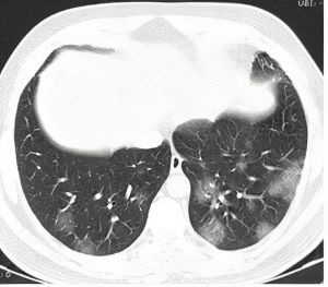
Purpose: To create a CT Score to objectively and quantitatively predict the severity and evolution of COVID-19 pneumonia in concordance with unenhanced upon admission chest CT findings. Material and Methods: We retrospectively evaluated 98 patients with a diagnosis of SARS-CoV-2 confirmed by RT-PCR admitted to the general ward. We developed a CT score to quantify the imaging involvement of the disease at hospital admission. This score values the type of patterns and the total burden of the lesion (expressed as a percentage of the parenchymal involvement). A Receiver Operating Characteristic (ROC) curve analysis was performed as a test of diagnostic accuracy of the developed Score. Results: 98 patients were analyzed, using as cut-off point a CT score puntaje ≤ 14. No patients with unfavourable evolution were detected (100 % Negative Predictive Value, 80 % sensitivity, 100 % specifity to predict favourable development). CT Score < 22 (91.2 % Negative Predictive Value for the need of oxygen reservoir masks and 94.7 % for unfavourable outcome). A CT Score ≥ 22 predicted a need for oxygen therapy and unfavourable development. (92.6 % Positive Predictive Value, 80 % sensitivity and 65 % specificity). The area under curve (AUC) was 0.8197, which makes it a test with a high diagnostic discriminatory capacity. Conclusion: CT Score is useful to determine the radiological assessment of pulmonary involvement in three grades: minor, moderate and severe. The imaging findings are highly correlated with clinical evolution variables. It can be considered an important tool for prognostic value and to adapt early and timely therapeutics behaviours in the development of this illness.
Downloads
References
Rubin G, Ryerson C, Haramati L, Sverzellati N, Kanne J, Raoof S, et al. The role of chest imaging in patient management during the COVID-19 pandemic: A multinatio nal consensus statement from the Fleischner Society. Radiology. 2020;296:172-80.
Simpson S, Kay F, AbbaraA, BhallaS, Chung J, Chung M, et al. Radiological Society of North America Expert Consensus Statement on Reporting Chest CT Findings Related to COVID-19. Endorsed by the Society of Thoracic Radiology, the American College of Radiology, and RSNA. Radiology. 2020;2(2).
Organización Mundial de la Salud. COVID-19; cronología de la actuación de la OMS [internet]. 2020, 27 de abril [citado: 2021 ene. 10]. Disponible en: https:// www.who.int/es/news/item/27-04-2020-who-timeline---covid-19
Pan F, Ye T, Sun P, Gui S, Liang B, Li L, et al. Time course of lung changes on chest CT during recovery from 2019 novel coronavirus (COVID-19) pneumonia. Radiology. 2020;200370
.Manli Wang, Ruiyuan Cao, Leike Zhang, Xinglou Yang, Jia Liu, Mingyue Xu; Zhengli Shi, Zhihong Hu, Wu Zhong, Gengfu Xial Remdesivir and chloroquine effectively inhibit the recently emerged novel coronavirus (2019-nCoV) in vitro. Cell Research (2020) 30:269–271; https://doi.org/10.1038/s41422-020-0282-0
Ramírez Cotrina CA. La radiología y pandemia COVID-19.Rev Per Radiol. 2020;20:1
Yuan M, Yin W, Tao Z, Tan W, Hu Y. Association of radiologic findings with morta lity of patients infected with 2019 novel coronavirus in Wuhan, China. PLoS ONE. 2020;15(3):e0230548. https://doi.org/10.1371/journal.pone.0230548
Chen N, Zhou M, Dong X, Qu J, Gong F, Han Y, et al. Epidemiological and clinical characteristics of 99 cases of 2019 novel coronavirus pneumonia in Wuhan, China: a descriptive study. Lancet. 2020.
Song F, Shi N, Shan F, Zhang Z, Shen J, et al. Novel coronavirus (2019-nCoV) Pneumonia. Radiology. 2020;295:210-7.
Dawei Wang, MD1; Bo Hu, MD1; Chang Hu, MD1; et. Clinical Characteristics of 138 Hospitalized Patients With 2019 Novel Coronavirus–Infected Pneumonia in Wuhan, China
WHO. Statement on the second meeting of the International Health Regulations (2005) Emergency Committee regarding the outbreak of novel coronavirus (2019- nCoV) [internet]. 2020, enero 30 [citado: 2021 ene, 20]. Disponible en: https://www. Who.Int/news-room/detail/30-01-2020-statement-on-thesecond-meeting-of-the-in ternational-health-regulations-(2005)-emergency-committee-regarding-theoutbreak of-novel-coronavirus-(2019-ncov)
Bernheim A, Mei X, Huang M, Yang Y, Fayad ZA, Zhang N, et al. Chest CT fin dings in coronavirus disease-19 (COVID-19): relationship to duration of infection. Radiology. 2020;20:200463.
Feng F, Jiang Y, Yuan M, Shen J, Yin H,Geng D, et al. Association of radiologic findings with mortality in patients with avian influenza H7N9 pneumonia. PloS one. 2014
Shi H, Han X,Zheng C. Evolution of CT manifestations in a patient recovered from 2019 novel coronavirus (2019-nCoV) pneumonia in Wuhan, China. Radiology. 2020:200269
Li K, Wu J, Wu F. The clinical and chest CT features associated with severe and critical COVID-19 pneumonia. Invest Radiol. 2020;55(6)
Downloads
Published
How to Cite
Issue
Section
License

This work is licensed under a Creative Commons Attribution-NonCommercial-ShareAlike 4.0 International License.
La Revista Colombiana de Radiología es de acceso abierto y todos sus artículos se encuentran libre y completamente disponibles en línea para todo público sin costo alguno.
Los derechos patrimoniales de autor de los textos y de las imágenes del artículo como han sido transferidos pertenecen a la Asociación Colombiana de Radiología (ACR). Por tanto para su reproducción es necesario solicitar permisos y se debe hacer referencia al artículo de la Revista Colombiana de Radiología en las presentaciones o artículos nuevos donde se incluyan.


