
Tridimensional late gadolinium enhancement at 3 T cardiac magnetic resonance for the evaluation of coronary venous anatomy: feasibility and findings
DOI:
https://doi.org/10.53903/01212095.156Keywords:
Coronary vessels, Cicatrix, Magnetic resonance imaging, Cardiac imaging techniquesAbstract
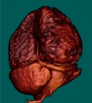
Objective: Knowledge of coronary venous anatomy (CVA) has critical importance for planning and performing electrophysiological procedures such as cardiac resynchronization therapy (CRT), left ventricle and right atrium ablation therapy and catheter arrhythmia mapping. Our aim is to evaluate the feasibility and applications of cardiac magnetic resonance imaging (RMC) performed at 3T for non-invasive depiction of CVA employing a three-dimensional late gadolinium enhancement sequence (3D RTG). Methods: 138 consecutive patients who underwent 3T RMC were evaluated using a 3D RTG sequence over a period of one year between 2016 and 2017. Identification of
different coronary venous structures as well as its relationship with myocardial fibrosis and other relevant clinical variables were recorded. Quality assessment was performed creating 3 groups (optimal, good, poor) according to visual assessment of each individual study. Association tests(Chi-square and Kruskall-Wallis) were performed. Results: The study included 62 women and
76 men with a median age of 48 (29-61) years. 3D RTG sequence yielded a diagnostic quality (optimal-good) for CVA evaluation in 76% of patients (p < 0.001). The following structures were identified (patients, %): anterior interventricular vein: 110 (79.7%), great cardiac vein: 109 (79%), posterior interventricular vein: 106 (76.8%), marginal vein: 53 patients (38.4%) and posterolateral vein: 74 (53.6%). Myocardial fibrosis was identified in 42 patients and epicardic fibrotic involvement of at least one coronary
vein path was recorded on 12% of patients on this subset. Shorter acquisition periods (p < 0.02) and performing the study under general anesthesia (p < 0.03) resulted in significantly better study quality. Conclusions: Non-invasive CVA evaluation is feasible with 3D RTG sequence obtained at 3T RMC. This approach may offer a valuable clinical tool for electrophysiological procedural planning.
Downloads
References
Blendea D, Shah RV, Auricchio A, Nandigam V, Orencole M, Heist EK, et al. Variability of coronary venous anatomy in patients undergoing cardiac resynchronization therapy: A high-speed rotational venography study. Hear Rhythm. 2007;4(9):1155-62.
Van de Veire NR, Marsan NA, Schuijf JD, Bleeker GB, Wijffels MCEF, van Erven L, et al. Noninvasive imaging of cardiac venous anatomy with 64-slice multi-slice computed tomography and noninvasive assessment of left ventricular dyssynchrony by 3-dimensional tissue synchronization imaging in patients with heart failure scheduled for cardiac re. Am J Cardiol. 2008;101(7):1023-9.
Tada H, Kurosaki K, Naito S, Koyama K, Itoi K, Ito S, et al. Three-dimensional visualization of the coronary venous system using multidetector row computed tomography. Circ J. 2005;69(2):165-70.
Echeverri D, Cabrales J, Jiménez A. Myocardial venous drainage: from anatomy to clinical use. J Invasive Cardiol. 2013;25(2):98-105.
Nguyên UC, Cluitmans MJM, Luermans JGLM, Strik M, de Vos CB, Kietselaer BLJH, et al. Visualisation of coronary venous anatomy by computed tomography angiography prior to cardiac resynchronisation therapy implantation. Netherlands Hear J. 2018;26(9):433-44.
Mischke K, Knackstedt C, Mühlenbruch G, Schimpf T, Neef P, Zarse M, et al. Imaging of the coronary venous system: Retrograde coronary sinus angiography versus venous phase coronary angiograms. Int J Cardiol. 2007;119(3):339-43.
Nguyên UC, Cluitmans MJM, Strik M, Luermans JG, Gommers S, Wildberger JE, et al. Integration of cardiac magnetic resonance imaging, electrocardiographic imaging, and coronary venous computed tomography angiography for guidance of left ventricular lead positioning. Europace. 2019;21(4):626-35. doi: 10.1093/europace/euy292.
Abbara S, Cury RC, Nieman K, Reddy V, Moselewski F, Schmidt S, et al. Noninvasive evaluation of cardiac veins with 16-MDCT angiography. AJR Am J Roentgenol. 2005;185(4):1001-6.
Mlynarski R, Mlynarska A, Sosnowski M. Anatomical variants of coronary venous system on cardiac computed tomography. Circ J. 2011;75(3):613-8. 10. Malagò R, Pezzato A, Barbiani C, Sala G, Zamboni GA, Tavella D, et al. Non invasive cardiac vein mapping: role of multislice CT coronary angiography. Eur J Radiol. 2012;81(11):3262-9.
Genc B, Solak A, Sahin N, Gur S, Kalaycioglu S, Ozturk V. Assessment of the coronary venous system by using cardiac CT. Diagnostic Interv Radiol. 2013;19(4):286-93.
Behar JM, Mountney P, Toth D, Reiml S, Panayiotou M, Brost A, et al. Real-Time X-MRI-guided left ventricular lead implantation for targeted delivery of cardiac resynchronization therapy. JACC Clin Electrophysiol. 2017;3(8):803-14.
Behar JM, Claridge S, Jackson T, Sieniewicz B, Porter B, Webb J, et al. The role of multi modality imaging in selecting patients and guiding lead placement for the delivery of cardiac resynchronization therapy. Expert Rev Cardiovasc Ther. 2017;15(2):93-107.
Bakos Z, Markstad H, Ostenfeld E, Carlsson M, Roijer A, Borgquist R. Combined preoperative information using a bullseye plot from speckle tracking echocardiography, cardiac CT scan, and MRI scan: targeted left ventricular lead implantation in patients receiving cardiac resynchronization therapy. Eur Heart J Cardiovasc Imaging. 2014;15(5):523-31.
Younger JF, Plein S, Crean A, Ball SG, Greenwood JP. Visualization of coronary venous anatomy by cardiovascular magnetic resonance. J Cardiovasc Magn Reson. 2009;11(1):26.
Nguyên UC, Mafi-Rad M, Aben J-P, Smulders MW, Engels EB, van Stipdonk AMW, et al. A novel approach for left ventricular lead placement in cardiac resynchronization therapy: Intraprocedural integration of coronary venous electroanatomic mapping with delayed enhancement cardiac magnetic resonance imaging. Hear Rhythm. 2017;14(1):110-9.
Shetty AK, Duckett SG, Ginks MR, Ma Y, Sohal M, Bostock J, et al. Cardiac magnetic resonance-derived anatomy, scar, and dyssynchrony fused with fluoroscopy to guide LV lead placement in cardiac resynchronization therapy: a comparison with acute haemodynamic measures and echocardiographic reverse remodelling. Eur Heart J Cardiovasc Imaging. 2013;14(7):692-9.
Kočková R, Sedláček K, Wichterle D, Šikula V, Tintěra J, Jansová H, et al. Cardiac resynchronization therapy guided by cardiac magnetic resonance imaging: A prospective, single-centre randomized study (RMC-CRT). Int J Cardiol. 2018;270:325-30.
Heydari B, Jerosch-Herold M, Kwong RY. Imaging for planning of cardiac resynchronization therapy. JACC Cardiovasc Imaging. 2012;5(1):93-110. 20. Chiribiri A, Kelle S, Götze S, Kriatselis C, Thouet T, Tangcharoen T, et al. Visualization of the cardiac venous system using cardiac magnetic resonance. Am J Cardiol. 2008;101(3):407-12.
Younger JF, Plein S, Crean A, Ball SG, Greenwood JP. Visualization of coronary venous anatomy by cardiovascular magnetic resonance. J Cardiovasc Magn Reson. 2009;11(1):26.
Khan FZ, Virdee MS, Palmer CR, Pugh PJ, O’Halloran D, Elsik M, et al. Targeted left ventricular lead placement to guide cardiac resynchronization therapy: the target study: a randomized, controlled trial. J Am Coll Cardiol. 2012;59(17):1509-18.
Jin Y, Zhang Q, Mao J, He B. Image-guided left ventricular lead placement in cardiac resynchronization therapy for patients with heart failure: a meta-analysis. BMC Cardiovasc Disord. 2015;15(1):36.
White JA, Yee R, Yuan X, Krahn A, Skanes A, Parker M, et al. Delayed enhancement magnetic resonance imaging predicts response to cardiac resynchronization therapy in patients with intraventricular dyssynchrony. J Am Coll Cardiol. 2006;48(10):1953-60.
Rathod RH, Powell AJ, Geva T. Myocardial fibrosis in congenital heart disease. Circ J. 2016;80(6):1300-7.
Downloads
Published
How to Cite
Issue
Section
License
Copyright (c) 2022 Revista Colombiana de Radiología

This work is licensed under a Creative Commons Attribution-NonCommercial-ShareAlike 4.0 International License.
La Revista Colombiana de Radiología es de acceso abierto y todos sus artículos se encuentran libre y completamente disponibles en línea para todo público sin costo alguno.
Los derechos patrimoniales de autor de los textos y de las imágenes del artículo como han sido transferidos pertenecen a la Asociación Colombiana de Radiología (ACR). Por tanto para su reproducción es necesario solicitar permisos y se debe hacer referencia al artículo de la Revista Colombiana de Radiología en las presentaciones o artículos nuevos donde se incluyan.


