
Pre-Surgical and Post-Surgical Assessment of the Upper Airway by Computed Tomography, In Patients with Obstructive Sleep Apnea Hypopnea Syndrome Undergoing Maxillomandibular Advancement Surgery
DOI:
https://doi.org/10.53903/01212095.3Keywords:
Palate, Sleep apnea syndromes, Mandibular advancement, TomographyAbstract
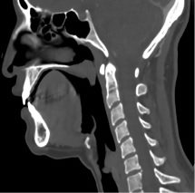
Introduction: Obstructive sleep apnea-hypopnea syndrome (OSAHS) secondary to craniofacial malformations increases morbidity and decreases quality of life of the patient. Objective: To describe the anatomical characteristics of the airway before and after surgery, in patients with OSAHS secondary to craniofacial malformations undergoing maxillo-mandibular advancement surgery. Methodology: This was a descriptive observational study in adults with OSAHS secondary to craniofacial malformations, attending the Clínica Universitaria Colombia, whom underwent maxillo-mandibular advancement surgery between December 2013 and October 2018. Pre-surgical and post-surgical measures at predefined anatomical repair points were analyzed, as well as the magnitude of the post-intervention change. Results: 57.17 % of the patients presented between 2 to 4 malformations, and the remaining 42.86 % presented 5 or more. The anatomical landmarks that presented significant changes in the post-surgical measures in subjects with 2 to 4 malformations were: The upper pharyngeal space 4.78 mm (SD 1.92), lower pharyngeal space 4.5 mm (SD 3.25), base of the epiglottis 3.21 mm (SD 2.80), the soft palate 4.92 mm (SD 2.84), the midpoint between the soft palate and the base of the epiglottis in its AP diameter 4.42 mm (SD 3.13) and transverse diameter 5.42 mm (SD 4.83). All patients with severe OSAHS presented post- surgical changes with statistically significant differences values that vary between 6.27 mm (p value 0.0011) and 4.72 mm (p value 0.0000). Conclusions: The changes in the pre- and post-surgical measures of airway anatomic landmark patients with OSAHS were characterized. These findings can be a guide for the radiologist at the time of reporting a study for this purpose, helping in the surgical planning and follow up of patients.
Downloads
References
Maurer J. Early diagnosis of sleep related breathing disorders portable monitoring. GMS Curr Top Otorhinolaryngol Head Neck Surg. 2008;7:1-20.
Lee W, Nagubadi S. Epidemiology of obstructive sleep apnea: a population-based perspective. Expert Rev Respir Med. 2008;349-64. https://doi.org/10.1586/17476348.2.3.349
Madani M. Reoperative treatment of obstructive sleep apnea. Oral Maxillofac Surg Clin NA. 2011;23(1):177-87. https://doi.org/10.1016/j.coms.2010.10.003
Sittitavornwong S, Waite PD. Imaging the upper airway in patients with sleep disor- dered breathing. Oral Maxillofac Surg Clin NA. 2009;21(4):389-402. https://doi.org/10.1016/j.coms.2009.08.004
Qaseem A, Holty J, Owens D, Dallas P, Starkey M, Shekelle P, et al. Management of obstructive sleep apnea in adults: A clinical practice guideline from the American College of Physicians. Ann Intern Med. 2013;159(7):471. https://doi.org/10.7326/0003-4819-159-7-201310010-00704
Won C, Li K, Guilleminault C. Surgical treatment of obstructive sleep apnea: Upper airway and maxillomandibular surgery. Proceed Am Thoracic Soc. 2008;5(2):193-9. https://doi.org/10.1513/pats.200708-121MG
Li H, Chen N-H, Wang C-R, Shu Y-H, Wang P-C. Use of 3-dimensional computed tomography scan to evaluate upper airway patency for patients undergoing sleep-disordered breathing surgery. Otolaryngol Head Neck Surg. 2003;129(4):336-42. https://doi.org/10.1016/S0194-5998(03)00629-6
Fairburn SC, Waite PD, Vilos G, Harding S, Bernreuter W, Cure J, et al. Three-dimen- sional changes in upper airways of patients with obstructive sleep apnea following maxillomandibular advancement. J Oral Maxillofac Surg. 2007;65(1):6-12. https://doi.org/10.1016/j.joms.2005.11.119
Faria AC, Nogueira da Silva-Junior S, García LV, dos Santos AC, França Fernandes MR, Veríssimo de Mello-Filho F. Volumetric analysis of the pharynx in patients with obstructive sleep apnea (OSA) treated with maxillomandibular advancement (MMA). Sleep Breath. 2013;17(1):395-401. https://doi.org/10.1007/s11325-012-0707-1
Abramson Z, Susarla S, Lawler M, Bouchard C, Troulis M, Kaban L. CT airway analysis and OSA. J Oral Maxillofac Surg. 2011;69(3):677-86. https://doi.org/10.1016/j.joms.2010.11.037
Hendler BH, Costello BJ, Silverstein K, Yen D, Goldberg A. A protocol for uvulo- palatopharyngoplasty, mortised genioplasty, and maxillomandibular advancement in patients with obstructive sleep apnea: An analysis of 40 cases. J Oral Maxillofac Surg. 2001;59(8)892-7. https://doi.org/10.1053/joms.2001.25275
Conley RS. Orthodontic considerations related to sleep- disorded breathing. Sleep Med Clin. 2010;5 (1):71-89. https://doi.org/10.1016/j.jsmc.2009.10.005
Vallejo-Balen A, Zabala-Parra SI, Amado S. Surgical treatment by otorhinolaryngology in obstructive sleep apnea-hypopnea syndrome. Rev. Fac. Med. 2017;65:109-14. https://doi.org/10.15446/revfacmed.v65n1Sup.59667
Downloads
Published
How to Cite
Issue
Section
License
Copyright (c) 2020 Revista Colombiana de Radiología

This work is licensed under a Creative Commons Attribution-NonCommercial-ShareAlike 4.0 International License.
La Revista Colombiana de Radiología es de acceso abierto y todos sus artículos se encuentran libre y completamente disponibles en línea para todo público sin costo alguno.
Los derechos patrimoniales de autor de los textos y de las imágenes del artículo como han sido transferidos pertenecen a la Asociación Colombiana de Radiología (ACR). Por tanto para su reproducción es necesario solicitar permisos y se debe hacer referencia al artículo de la Revista Colombiana de Radiología en las presentaciones o artículos nuevos donde se incluyan.


