
Oncocytomas: Imaging Findings and Anatomopathology Correlation
DOI:
https://doi.org/10.53903/01212095.42Keywords:
Oncocitoma, Neoplasias renales, Diagnóstico, Tomografía computarizada multidetectorAbstract
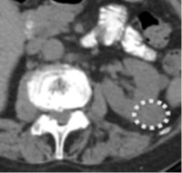
Introduction: The imaging findings of oncocytomas usually coincide with renal cell carcinoma (RCC), which makes it difficult to discriminate them in imaging. Objective: To evaluate the imaging findings of a sample of oncocytomas in tomography (CT). Methods: We retrospectively selected patients with renal tumor surgery and oncocytoma anatomopathological diagnosis, who were treated between January 2015 and December 2017. Patients who underwent CT with intravenous contrast at our institution were included. Results: Of the total number of patients (n = 44), 43 had a single renal lesion while one patient presented 3 lesions. Of the total lesions (n = 47), 20 (42.55%) were diagnosed after a radical nephrectomy and 24 (51.10%) were diagnosed by a partial nephrectomy. The mean maximum diameter was 36.5 mm (RIQ 22-44, 25), of which they were grouped by tumor length into smaller or larger than 4 cm, with 22 tumors in this last group (47%). Of these, 15 tumors (31.91 %) that were larger than 4 cm had a central scar. Calcifications were evident in 3 patients (6.8 %). One tumor (2.1%) was found with the presence of inversion of segmental enhancement after administration of intravenous contrast. In this case, the oncocytoma was less than 4 cm. Conclusion: The finding of a solid mass with more enhancement than the surrounding parenchyma during the nephrographic phase makes it necessary to consider oncocytoma among the differential diagnoses
Downloads
References
Woo S, Cho JY, Kim SH, Kim SY, Lee HJ, Hwang SI, et al. Segmental enhancement inversion of small renal oncocytoma: differences in prevalence according to tumor size. AJR Am J Roentgenol. 2013;200(5):1054-9.
Gakis G, Kramer U, Schilling D, Kruck S, Stenzl A, Schlemmer H-P. Small renal oncocytomas: Differentiation with multiphase CT. Eur J Radiol. 2011;80:274-8. http:// dx.doi.org/10.1016/j.ejrad.2010.06.049.
Davidson AJ, Hayes WS, Hartman DS, McCarthy WF, Davis CJ. Renal oncocytoma and carcinoma: failure of differentiation with CT. Radiology. 1993;186:693-6. http:// dx.doi.org/10.1148/radiology.186.3.8430176.
Chao DH, Zisman A, Pantuck AJ, Freedland SJ, Said JW, Belldegrun AS. Changing concepts in the management of renal oncocytoma. Urology. 2002;59(5):635-42.
Duchene DA, Lotan Y, Cadeddu JA, Sagalowsky AI, Koeneman KS. Histopathology of surgically managed renal tumors: analysis of a contemporary series. Urology. 2003;62:827-30. http://dx.doi.org/10.1016/s0090-4295(03)00658-7.
Filipas D, Fichtner J, Spix C, Black P, Carus W, Hohenfellner R, et al. Nephron sparing surgery of renal cell carcinoma with a normal opposite kidney: long-term . outcome in 180 patients. Urology. 2000;56:387-92. http://dx.doi.org/10.1016/s0090-4295(00)00656-7.
Frank I, Blute ML, Cheville JC, Lohse CM, Weaver AL, Zincke H. Solid renal tu mors: an analysis of pathological features related to tumor size. J Urol. 2003;170(6 Pt 1):2217-20.
Israel GM, Bosniak MA. How I Do It: Evaluating renal masses. Radiology. 2005;236:441-50. http://dx.doi.org/10.1148/radiol.2362040218.
Stakhovskyi O, Yap SA, Leveridge M, Lawrentschuk N, Jewett MAS. Small renal mass: what the urologist needs to know for treatment planning and assessment of treatment results. AJR Am J Roentgenol. 2011;196(6):1267-73.
Bird VG, Kanagarajah P, Morillo G, Caruso DJ, Ayyathurai R, Leveillee R, et al. Differentiation of oncocytoma and renal cell carcinoma in small renal masses (<4 cm): the role of 4-phase computerized tomography. World J Urol. 2011;29(6):787-92.
Pierorazio PM, Hyams ES, Tsai S, Feng Z, Trock BJ, Mullins JK, et al. Multiphasic enhancement patterns of small renal masses (≤4 cm) on preoperative computed tomo graphy: utility for distinguishing subtypes of renal cell carcinoma, angiomyolipoma, and oncocytoma. Urology. 2013;81(6):1265-71.
Zhang J, Lefkowitz RA, Ishill NM, Wang L, Moskowitz CS, Russo P, et al. Solid renal cortical tumors: differentiation with CT. Radiology. 2007;244(2):494-504.
Kim JI, Cho JY, Moon KC, Lee HJ, Kim SH. Segmental enhancement inversion at biphasic multidetector CT: characteristic finding of small renal oncocytoma. Radiology. 2009;252(2):441-8.
McGahan JP, Lamba R, Fisher J, Starshak P, Ramsamooj R, Fitzgerald E, et al. Is Segmental Enhancement Inversion on Enhanced Biphasic MDCT a reliable sign for the noninvasive diagnosis of renal oncocytomas? Am J Roentgenol. 2011;197:W674-9. http://dx.doi.org/10.2214/ajr.11.6463.
Schieda N, McInnes MDF, Cao L. Diagnostic accuracy of segmental enhancement inversion for diagnosis of renal oncocytoma at biphasic contrast enhanced CT: sys tematic review. Eur Radiol. 2014;24(6):1421-9.
O’Malley ME, Tran P, Hanbidge A, Rogalla P. Small renal oncocytomas: is segmental enhancement inversion a characteristic finding at biphasic MDCT? AJR Am J Roent genol. 2012;199(6):1312-5.
Sasaguri K, Takahashi N, Gómez-Cardona D, Leng S, Schmit GD, Carter RE, et al. Small (< 4 cm) Renal Mass: Differentiation of Oncocytoma from renal cell carcinoma on biphasic contrast-enhanced CT. AJR Am J Roentgenol. 2015;205(5):999-1007.
Galia M, Albano D, Bruno A, Agrusa A, Romano G, Di Buono G, et al. Imaging features of solid renal masses. Br J Radiol. 2017;90(1077):20170077.
Kuroda N, Toi M, Yamamoto M, Miyazaki E, Hayashi Y, Hiroi M, et al. Immuno histochemical identification of intracytoplasmic lumens by cytokeratin typing may differentiate renal oncocytomas from chromophobe renal cell carcinomas. Histol Histopathol. 2004;19(1):23-8.
Beland MD, Mayo-Smith WW, Dupuy DE, Cronan JJ, DeLellis RA. Diagnostic yield of 58 consecutive imaging-guided biopsies of solid renal masses: should we biopsy all that are indeterminate? AJR Am J Roentgenol. 2007;188(3):792-7.
Heilbrun ME, Zagoria RJ, Julian Garvin A, Craig Hall M, Krehbiel K, Southwick A, et al. CT-Guided biopsy for the diagnosis of renal tumors before treatment with percutaneous ablation. Am J Roentgenol. 2007;188:1500-5. http://dx.doi.org/10.2214/ajr.06.0389.
Gillies RJ, Kinahan PE, Hricak H. Radiomics: Images are more than pictures, they are data. Radiology. 2016;278(2):563-77
Downloads
Published
How to Cite
Issue
Section
License
Copyright (c) 2021 Revista Colombiana de Radiología

This work is licensed under a Creative Commons Attribution-NonCommercial-ShareAlike 4.0 International License.
La Revista Colombiana de Radiología es de acceso abierto y todos sus artículos se encuentran libre y completamente disponibles en línea para todo público sin costo alguno.
Los derechos patrimoniales de autor de los textos y de las imágenes del artículo como han sido transferidos pertenecen a la Asociación Colombiana de Radiología (ACR). Por tanto para su reproducción es necesario solicitar permisos y se debe hacer referencia al artículo de la Revista Colombiana de Radiología en las presentaciones o artículos nuevos donde se incluyan.


