
Intravessical Fat-Fluid Level Secondary to Silent Bladder Perforation After Transurethral Resection of Urothelial Carcinoma
DOI:
https://doi.org/10.53903/01212095.44Keywords:
Urinary bladder, Carcinoma transitional cell Ultrasonography, Multidetector computed tomographyAbstract
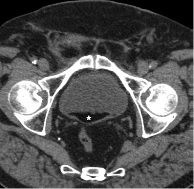
Non-muscle-invasive bladder tumours are defined as papillary lesions limited to the bladder mucosa or invading the lamina propia, in addition to flat morphology and high-grade tumors limited to the mucosa (carcinoma in situ [CIS]). Diagnostic confirmation occurs after histological analysis of the sample obtained in the transurethral resection of the bladder (TURB), which in Ta-T1 tumours requires complete resection of all lesions, including part of the detrusor muscle, being in these cases also the therapeutic method. In the case of CIS, which may simulate inflammation or not be visible in cystoscopy, multiple random bladder biopsies are necessary (2). Iatrogenic bladder perforation is the second most frequent adverse event of this procedure; This solution of continuity in the wall can lead to the migration of fat into the bladder, which in imaging tests results in the formation of an intravesical fat-fluid level, a rare finding, which in turn has a broad differential diagnosis that covers primary, infectious and traumatic causes, but not previously described as a complication of TURB. We report the case of an asymptomatic patient with presence of intravesical fat-fluid, secondary to extra-peritoneal bladder perforation, after transurethral resection of a non-muscle[1]invasive urothelial carcinoma in the bladder dome.
Downloads
References
Brierley JD, Gospodarowicz MK, Wittekind C. TNM classification of malignant tumours. 8th ed. Oxford: Wiley-Blackwell; 2017.
Babjuk M, Böhle A, Burger M, Capoun O, Cohen D, Compérat EM, et al. EAU guidelines on non-muscle-invasive urothelial carcinoma of the bladder: Update 2016. Eur Urol. 2017;71:447-61.
Collado A, Cechile GE, Salvador J, Vicente J. Early complications of endoscopic treatment for superficial bladder tumors. J Urol. 2000;164:1529-32.
Summerton DJ, Kitrey ND, Lumen N, Serafetinidis E, Djakovic N. European Asso ciation of Urology. EAU guidelines on iatrogenic trauma. Eur Urol. 2012;62:628-39.
Balbay MD, Cimentepe E, Unsal A, Bayrak O, Koc A, Akbulut Z. The actual incidence of bladder perforation following transurethral bladder surgery. J Urol. 2005;174:2260-2.
Vaccaro JP, Brody JM. CT cystography in the evaluation of major bladder trauma. Radiographics. 2000;20:1373-81.
Lim AK, Johns Putra LG, Troy AJ, Ierino FL. Intravesical fat entrapment as a cause of failure of extraperitoneal bladder perforation to heal spontaneously. Int Urol Nephrol. 2007;39:795-8.
Chan DP, Abujudeh HH, Cushing GL Jr, Novelline RA. CT cystography with multi planar reformation for suspected bladder rupture: experience in 234 cases. AJR Am J Roentgenol. 2006;187:1296-302.
Chow LC, Kwan SW, Olcott EW, Sommer G. Split-bolus MDCT Urography with synchronous nephrographic and excretory phase enhancement. AJR Am J Roentgenol. 2007;189:314-22.
Tan Y. Chyluria in non-filarial endemic areas: an internist’s perspective. BMJ Case Reports. 2017;2017:bcr-2017-220772.
Downloads
Published
How to Cite
Issue
Section
License

This work is licensed under a Creative Commons Attribution-NonCommercial-ShareAlike 4.0 International License.
La Revista Colombiana de Radiología es de acceso abierto y todos sus artículos se encuentran libre y completamente disponibles en línea para todo público sin costo alguno.
Los derechos patrimoniales de autor de los textos y de las imágenes del artículo como han sido transferidos pertenecen a la Asociación Colombiana de Radiología (ACR). Por tanto para su reproducción es necesario solicitar permisos y se debe hacer referencia al artículo de la Revista Colombiana de Radiología en las presentaciones o artículos nuevos donde se incluyan.


