
Multinodular and Vacuolating Posterior Fossa Lesions of Unknown Significance. Case Report in Colombia
DOI:
https://doi.org/10.53903/01212095.45Keywords:
Magnetic resonance imaging, Brain neoplasm, CerebellumAbstract
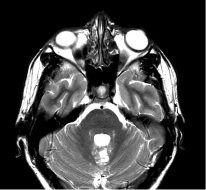
There are multiple types of cystic lesions that can be found in the brain and in the posterior fossa. Among these, a new entity called MV PLUS (Multinodular and Vacuolating Posterior Fossa Lesions of Unknown Significance) has similar imaging characteristics to the vacuolating multinodular tumor, but as indicated by its acronym in English, it is located in the posterior fossa. These tumors are defined as a group of small, high intensity nodular subcortical images in the T2-FLAIR, with or without post-contrast enhancement. They can be differentiated from other cystic entities, because they are clustered lesions, without reactive inflammatory changes, in a cortex of normal appearance and without changes at follow-up. The latter, causes them to be considered benign, non-aggressive lesions. We present the case of a 21-year-old patient, who presented an incidentally found lesion in the central region of the vermis. MRI showed a multicystic-looking mass, that had no changes at follow-up in the last 3 years and without obvious differential diagnosis, suggestive of MV PLUS.
Downloads
References
Nunes RH, Hsu CC, da Rocha AJ, do Amaral LLF, Godoy LFS, Watkins TW, et al. Multinodular and vacuolating neuronal tumor of the cerebrum: A new “Leave Me Alone” lesion with a characteristic imaging pattern. Am J Neuroradiol. 2017;38(10):1899-904.
Lecler A, Bailleux J, Carsin B, Adle-Biassette H, Baloglu S, Bogey C, et al. Multinodular and vacuolating posterior fossa lesions of unknown significance. Am J Neuroradiol. 2019;40(10):1689-94.
Yamaguchi M, Komori T, Nakata Y, Yagishita A, Morino M, Isozaki E. Multinodular and vacuolating neuronal tumor affecting amygdala and hippocampus: A quasi-tumor? Multinodular and vacuolating neuronal tumor. Pathol Int. 2016;66(1):34-41.
Shin JH, Lee HK, Khang SK, Kim DW, Jeong AK, Ahn KJ, et al. Neuronal tumors of the central nervous system: Radiologic findings and pathologic correlation. Radio- Graphics. 2002;22(5):1177-89.
Shih RY, Koeller KK. Bacterial, fungal, and parasitic infections of the central nervous system: Radiologic-pathologic correlation and historical perspectives: From the Radiologic Pathology Archives. RadioGraphics. 2015;35(4):1141-69.
Huse JT, Edgar M, Halliday J, Mikolaenko I, Lavi E, Rosenblum MK. Multinodular and vacuolating neuronal tumors of the cerebrum: 10 cases of a distinctive seizureassociated lesion: multinodular and vacuolating neuronal tumors. Brain Pathol. 2013;23(5):515-24.
Sutton LN. Cerebellar astrocytomas [internet]. Clinical Gate. 202AD. Disponible en: https://clinicalgate.com/cerebellar-astrocytomas/
Van der Knaap MS, Smit LME, Barth PG, Catsman-Berrevoets CE, Brouwer OF, Begeer JH, et al. Magnetic resonance imaging in classification of congenital muscular dystrophies with brain abnormalities. Ann Neurol. 1997;42(1):50-9.
Downloads
Published
How to Cite
Issue
Section
License

This work is licensed under a Creative Commons Attribution-NonCommercial-ShareAlike 4.0 International License.
La Revista Colombiana de Radiología es de acceso abierto y todos sus artículos se encuentran libre y completamente disponibles en línea para todo público sin costo alguno.
Los derechos patrimoniales de autor de los textos y de las imágenes del artículo como han sido transferidos pertenecen a la Asociación Colombiana de Radiología (ACR). Por tanto para su reproducción es necesario solicitar permisos y se debe hacer referencia al artículo de la Revista Colombiana de Radiología en las presentaciones o artículos nuevos donde se incluyan.


