
Usefulness of 3D Digital Subtraction Angiography in the Endovascular Approach of Cerebral Aneurysms
DOI:
https://doi.org/10.53903/01212095.53Keywords:
Digital subtraction angiography, Intracranial aneurysm, Embolization, therapeutic, Endovascular proceduresAbstract
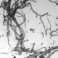
Introduction: The trend in management of intracranial aneurysms has shifted during the last decades to minimally invasive endovascular procedures. The usefulness of new imaging tools such as digital subtraction angiography in 3D (3D DSA), added to the experience of neurointerventional radiologists, have led to greater definition and accuracy in the study of intracranial aneurysms. Objective: To describe the usefulness of three-dimensional digital subtraction angiography for pre and post embolization approach of intracranial aneurysms. Methodology: A cross-sectional study between January 2016 and April 2017 in patients diagnosed with arterial cerebral aneurysms at the Hospital Infantil Universitario San José in Bogota, Colombia. Results: 32 patients were included, 71.8% (n = 23) were women. Among the risk factors for aneurysm rupture, the most frequent was age above 40 years (81.8%). The most frequent location was in the Right Middle Cerebral Artery (MCA) (30.3%). All cases corresponded to saccular aneurysms. In the immediate post-embolization angiographic control it was evidence that 16 cases (48.5%) presented residual sac. Conclusions: The realization of multiplanar projections with 3D angiographic reconstruction allows for a better characterization of the aneurysm and evaluation of the adjacent anatomical structures, being very useful for the planning of the procedure and in the follow-up.
Downloads
References
Molyneux AJ, Kerr RSC, Yu L-M, et al. International subarachnoid aneurysm trial (ISAT) of neurosurgical clipping versus endovascular coiling in 2143 patients with ruptured intracranial aneurysms: a randomised comparison of effects on survival, dependency, seizures, rebleeding, subgroups, and aneurysm occlusion. Lancet (London, England). 2005;366(9488):809-17.
Cieściński J, Serafin Z, Strześniewski P, et al. DSA volumetric 3D reconstructions of intracranial aneurysms: A pictorial essay. Polish J Radiol. 2012;77(2):47-53.
Anxionnat R, Bracard S, Ducrocq X, et al. Intracranial aneurysms: Clinical Value of 3D digital subtraction angiography in the therapeutic decision and endovascular treatment. Radiology. 2001;218(3):799-808.
Tanoue S, Kiyosue H, Kenai H, et al. Three-dimensional reconstructed images after rotational angiography in the evaluation of intracranial aneurysms: Surgical correlation. Neurosurgery. 2000;47(4):866-71.
Robbins SL, Kumar V, Cotran RS. Robbins and Cotran pathologic basis of disease. 8th ed. Philadelphia PA: Saunders/Elsevier; 2010.
Zhou B, Li M-H, Wang W, et al. Three-dimensional volume-rendering technique in the angiographic follow-up of intracranial aneurysms embolized with coils. J Neurosurg. 2010;112(3):674-80.
Hacein-Bey L, Provenzale JM. Current imaging assessment and treatment of intracranial aneurysms. Am J Roentgenol. 2011;196(1):32-44.
Grobelny TJ. Brain aneurysms: Epidemiology, treatment options, and milestones of endovascular treatment evolution. Disease-a-Month. 2011;57(10):647-55.
Fifi JT, Meyers PM, Lavine SD, et al. Complications of modern diagnostic cerebral angiography in an Academic Medical Center. JVIR. 2009;20:442-7.
Bau Alegría J. Reconstrucción 3D angiográfica en el diagnóstico y el tratamiento de aneurismas cerebrales. Imagen Diagnóstica. 2010;1(2):51-5.
Sugahara T, Korogi Y, Nakashima K, et al. Comparison of 2D and 3D digital subtraction angiography in evaluation of intracranial aneurysms. AJNR Am J Neuroradiol. 2002;23(9):1545-52.
Van Rooij WJ, Sprengers ME, de Gast AN, et al. 3D Rotational angiography: The new gold standard in the detection of additional intracranial aneurysms. Am J Neuroradiol. 2008;29(5):976-9.
Hochmuth A, Spetzger U, Schumacher M. Comparison of three-dimensional rotational angiography with digital subtraction angiography in the assessment of ruptured cerebral aneurysms. Am J Neuroradiol. 2002;23(7):1199-205.
Downloads
Published
How to Cite
Issue
Section
License

This work is licensed under a Creative Commons Attribution-NonCommercial-ShareAlike 4.0 International License.
La Revista Colombiana de Radiología es de acceso abierto y todos sus artículos se encuentran libre y completamente disponibles en línea para todo público sin costo alguno.
Los derechos patrimoniales de autor de los textos y de las imágenes del artículo como han sido transferidos pertenecen a la Asociación Colombiana de Radiología (ACR). Por tanto para su reproducción es necesario solicitar permisos y se debe hacer referencia al artículo de la Revista Colombiana de Radiología en las presentaciones o artículos nuevos donde se incluyan.


