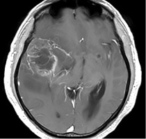
Gliosarcoma: Case Report and Findings by Tomography and Conventional and DTI Magnetic Resonance Imaging
DOI:
https://doi.org/10.53903/01212095.55Keywords:
Gliosarcoma, Tomography, X-ray computed, Magnetic resonance imagingAbstract
Gliosarcoma is a rare and highly malignant central nervous system tumor. It is classified by the WHO as a variant of glioblastoma (grade IV) and has a poor prognosis. Histologically it is characterized by having both glial and mesenchymal components. Clinically, it varies depending on the location and size of the tumor, the most frequent symptoms being seizures, headaches and focal neurological deficit. The initial diagnostic approach is computed tomography, which provides suspicionus data; however, magnetic resonance is the diagnostic pillar, providing important data that becomes more significant with the use of functional sequences such as tractography. A clinical case is presented with a literature review and the most significant findings in the imaging studies
Downloads
References
De Robles P, Fiest KM, Frolkis AD, Pringsheim T, Atta C, St Germaine-Smith C, et al. The worldwide incidence and prevalence of primary brain tumors: a systematic review and meta-analysis. Neuro Oncol. 2015;17(6):776-83.
Boerman RH, Anderl K, Herath J, et al. The glial and mesenchymal elements of glio sarcomas share similar genetic alterations. J Neuropathol Exp Neurol. 1996;55:973-81.
Kakkar N, Kaur J, Singh G, Singh P, Siraj F, Gupta A. Gliosarcoma in young adults: A rare variant of glioblastoma. W J Oncology. 2017;8(2):53-7.
Maiuri F, Stella L, Benvenuti D, et al. Cerebral gliosarcomas: correlation of computed tomographic findings, surgical aspect, pathological features, and prognosis. Neuro surgery. 1990;26:261-7.
Anderson MD, Colen RR, Tremont-Lukats IW. Imaging mimics of primary malignant tumors of the central nervous system (CNS). Curr Oncol Rep. 2014;16:399.
Chourmouzi D, Papadopoulou E, Marias K, Drevelegas A. Imaging of brain tumors. Surg Oncol Clin N Am. 2014;23(4):629-84.
Burtscher IM, Skagerberg G, Geijer B, Englund E, Ståhlberg F, Holtås S. Proton MR spectroscopy and preoperative diagnostic accuracy: an evaluation of intracranial mass lesions characterized by stereotactic biopsy findings. AJNR Am J Neuroradiol. 2000;21:84-93.
Ma J, Su S, Yue S, Zhao Y, Li Y, Chen X, Ma H, et al. Preoperative visualization of cranial nerves in skull base tumor surgery using diffusion tensor imaging technology. Turk Neurosurg. 2016;26(6):805-12.
WI, Zhang F, Unadkat P, et al. White matter tractography for neurosurgical planning: A topography-based review of the current state of the art. Essayed NeuroImage: Clin. 2017;15:659-72.
Wen PY, Macdonald DR, Reardon DA, Cloughesy TF, Sorensen AG, Galanis E, Degroot J, Wick W, Gilbert MR, Lassman AB, Tsien C, Mikkelsen T, Wong ET, Chamberlain MC, Stupp R, Lamborn KR, Vogelbaum MA, van den Bent MJ, Chang SM. Updated response assessment criteria for high-grade gliomas: response assessment in neuro-oncology working group. J Clin Oncol. 2010;28(11):1963-72.
Hygino da Cruz LC Jr, Rodríguez I, Dominguez RC, Gasparetto EL, Sorensen AG. Pseudoprogression and pseudoresponse: imaging challenges in the assessment of posttreatment glioma. AJNR Am J Neuroradiol. 2011; 32(11):1978-85.

Downloads
Published
How to Cite
Issue
Section
License

This work is licensed under a Creative Commons Attribution-NonCommercial-ShareAlike 4.0 International License.
La Revista Colombiana de Radiología es de acceso abierto y todos sus artículos se encuentran libre y completamente disponibles en línea para todo público sin costo alguno.
Los derechos patrimoniales de autor de los textos y de las imágenes del artículo como han sido transferidos pertenecen a la Asociación Colombiana de Radiología (ACR). Por tanto para su reproducción es necesario solicitar permisos y se debe hacer referencia al artículo de la Revista Colombiana de Radiología en las presentaciones o artículos nuevos donde se incluyan.


