
Recurrent Pneumonia as a Manifestation of Pulmonary Sequestration. Case Report
DOI:
https://doi.org/10.53903/01212095.56Keywords:
Bronchopulmonary sequestration, Pneumonia, Congenital anomalies, Multidetector computed tomographyAbstract
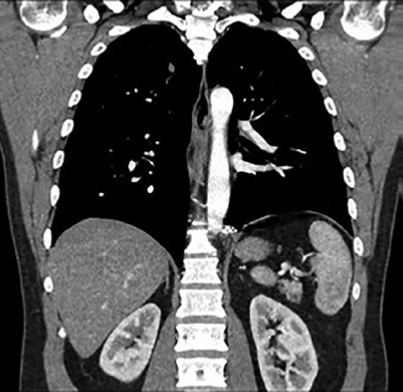
Pulmonary sequestration is a congenital respiratory disease that involves the pulmonary parenchymaand its vasculature. It can be divided into intralobar or extralobar depending on its relationshipwith normal visceral pleura. The extralobar subtype is usually diagnosed prenatally or in the firstmonths of life, while the intralobar occurs in young adults. It represents approximately 0.15-6.4%of all congenital lung malformations and does not show differences with respect to sex with a 1:1ratio. We present the case of an average adult male patient with recurrent pneumonia in whommultiples imaging studies are performed, with tomographic findings compatible with intralobarpulmonary sequestration in the left posterior basal segment, with the aim of performing a literaturereview to raise a discussion on the importance of the proper and early diagnosis of this pathology.It is necessary to have a high clinical suspicion -it is a disease of variable presentation-, to requestthe most appropriate imaging study, since the images are those that give the definitive diagnosisof pulmonary sequestration. In some cases, management can be performed in a minimally invasivemanner by interventional radiology, as in this case.
Downloads
References
Correia-Pinto J, Gonzaga S, Huang Y, Rottier R. Congenital lung lesions—underlying molecular mechanisms. Semin Pediatr Surg. 2010;19(3):171-9.
Newman B. Congenital bronchopulmonary foregut malformations: concepts and controversies. Pediatr Radiol. 2006;36(8):773-91.
Savic B, Birtel FJ, Tholen W, Funke HD, Knoche R. Lung sequestration: report of seven cases and review of 540 published cases. Thorax. 1979;34:96-101.
Stocker JT, Kagan-Hallet K. Extralobar pulmonary sequestration: analysis of 15 cases. Am J Clin Pathol. 1979;72(6):917-25.
Rosado de Christenson ML, Frazier AA, Stocker JT, Templeton PA. Extralobar sequestration: radiologic-pathologic correlation. Radiographics. 1993;13:425-41.
Long Q, Zha Y, Yang Z. Evaluation of pulmonary sequestration with multidetector computed tomography angiography in a select cohort of patients: A retrospective study. Clinics (Sao Paulo). 2016;71:392-8.
Felker RE, Tonkin IL. Imaging of pulmonary sequestration. AJR Am J Roentgenol. 1990;154(2):241-9.
Stocker JT, Malczak HT. A study of pulmonary ligament arteries: relationship to intralobar pulmonary sequestration. Chest. 1984;86:611-5.
Laberge JM, Bratu I, Flageole H. The management of asymptomatic congenital lung malformations. Paediatr Respir Rev. 2004;5:305-12.
Belchis D, Cowan M, Mortman M, Rezvani B. Adenocarcinoma arising in an ex- tralobar sequestration: a case report and review of the literatura. Lung Cancer. 2014;84:92-5.
Downloads
Published
How to Cite
Issue
Section
License

This work is licensed under a Creative Commons Attribution-NonCommercial-ShareAlike 4.0 International License.
La Revista Colombiana de Radiología es de acceso abierto y todos sus artículos se encuentran libre y completamente disponibles en línea para todo público sin costo alguno.
Los derechos patrimoniales de autor de los textos y de las imágenes del artículo como han sido transferidos pertenecen a la Asociación Colombiana de Radiología (ACR). Por tanto para su reproducción es necesario solicitar permisos y se debe hacer referencia al artículo de la Revista Colombiana de Radiología en las presentaciones o artículos nuevos donde se incluyan.


