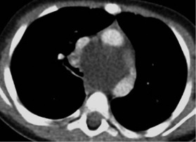
Radiological Manifestations of Congenital Lung Malformations. Experience of three Hospitals in Bogotá
DOI:
https://doi.org/10.53903/01212095.67Keywords:
Congenital abnormalities, Multidetector computed tomography, Lung diseases, ChildAbstract
Objective: To describe the radiological characteristics of congenital pulmonary and airway malformations which are frequently found in pediatric patients, from three hospitals in Bogotá between the years 2010 - 2016. Materials and methods: Retrospective, observational and descriptive study with a sample of 27 patients, with an average age of 5 months, who met inclusion criteria: patients between 0 months and 17 years of age, with a confirmed diagnosis of congenital malformation of the lung, who underwent surgery for lung or airway lesion and whose histopathological study was compatible with congenital malformation of the lung. Results: The prevalence of congenital malformations is higher in females. 80% of cases had prenatal diagnosis, with cystic adenomatoid malformation being the most common and the main radiological feature being the cyst. Conclusion: Computed tomography allows detailed studies of these malformations, achieving greater accuracy compared to conventional techniques such as chest radiography and ultrasound.
Downloads
References
Biyyam DR, Chapman T, Ferguson MR, Deutsch G, Dighe MK. Congenital lung abnormalities: Embryologic features, prenatal diagnosis, and postnatal radiologicpathologic correlation. RadioGraphics. 2010;30(6):1721-38.
Lee EY, Dorkin H, Vargas SO. Congenital pulmonary malformations in pediatric patients: review and update on etiology, classification, and imaging findings. Radiol Clin North Am. 2011;49(5):921-48.
Lee EY, Boiselle PM, Cleveland RH. Multidetector CT evaluation of congenital lung anomalies 1. Radiology. 2008;247(3):632-48.
Antón-Martín P, Cuesta-Rubio MT, López-González MF, Ortiz-Movilla R, Lorente- Jareño ML, López-Rodríguez E, et al. Malformación adenomatoidea quística congénita. Rev chil. pediatría. 2011;82:129-36.
Ch’In KY, Tang MY. Congenital adenomatoid malformation of one lobe of a lung with general anasarca. Arch Pathol (Chic). 1949;48(3):221-9.
Stocker JT, Madewell JE, Drake RM. Congenital cystic adenomatoid malformation of the lung. Classification and morphologic spectrum. Hum Pathol. 1977;8(2):155-71.
Stocker JT. Congenital pulmonary airway malformation: A new name and an expanded classification of congenital cystic adenomatoid malformation of the lung. Histopathology. 2002;41(Suppl 2):424-31.
Stocker JT. Cystic lung disease in infants and children. Fetal Pediatr Pathol. 2009;28(4):155-84.
Mehta AA, Viswanathan N, Vasudevan AK, Paulose R, Abraham M. Congenital cystic adenomatoid malformation: A tertiary care hospital experience. J Clin Diagn Res. 2016;10(11):SC01-SC4.
Trotman-Dickenson B. Congenital lung disease in the adult: guide to the evaluation and management. J Thorac Imaging. 2015;30(1):46-59.
Kao SW, Zuppan CW, Young LW. AIRP Best cases in radiologic-pathologic correlation: Type 2 congenital cystic adenomatoid malformation (Type 2 congenital pulmonary airway malformation). RadioGraphics. 2011;31(3):743-8.
Williams HJ, Johnson KJ. Imaging of congenital cystic lung lesions. Paediatr Respir Rev. 2002;3(2):120-7.
Tashtoush B, Memarpour R, González J, Gleason JB, Hadeh A. Pulmonary sequestration: A 29 patient case series and review. J Clin Diagn Res. 2015;9(12):Ac05-8.
Pryce DM. Lower accessory pulmonary artery with intralobar sequestration of lung; a report of seven cases. J Pathol Bacteriol. 1946;58(3):457-67.
Clements BS, Warner JO. Pulmonary sequestration and related congenital bronchopulmonary- vascular malformations: nomenclature and classification based on anatomical and embryological considerations. Thorax. 1987;42(6):401-8.
Wei Y, Li F. Pulmonary sequestration: a retrospective analysis of 2625 cases in China. Eur J Cardiothorac Surg. 2011;40(1):e39-42.
Qian X, Sun Y, Liu D, Wu X, Wang Z, Tang Y. Pulmonary sequestration: a case report and literature review. Int J Clin Exp Med. 2015;8(11):21822-5.
Berrocal T, Madrid C, Novo S, Gutiérrez J, Arjonilla A, Gómez-León N. Congenital anomalies of the tracheobronchial tree, lung, and mediastinum: Embryology, radiology, and pathology. RadioGraphics. 2004;24(1):e17-e. Pubmed.
Heithoff KB, Sane SM, Williams HJ, Jarvis CJ, Carter J, Kane P, et al. Bronchopulmonary foregut malformations. A unifying etiological concept. AJR Am J Roentgenol. 1976;126(1):46-55.
Jeung MY, Gasser B, Gangi A, Bogorin A, Charneau D, Wihlm JM, et al. Imaging of cystic masses of the mediastinum. Radiographics. 2002;22(Spec No):S79-93.
Kaistha A, Levine J. An unusual cause of pediatric dysphagia: Bronchogenic cyst. Glob Pediatr Health. 2017;4:2333794x16686492.
Rogers LF, Osmer JC. Bronchogenic cyst. A review of 46 cases. Am J Roentgenol Radium Ther Nucl Med. 1964;91:273-90.
Abdellah O, Mohamed H, Youssef B, Abdelhak B. A case of congenital lobar emphysema in the middle lobe. J Clin Neonatol. 2013;2(3):135-7.
Latif I, Shamim S, Ali S. Congenital lobar emphysema. J Pak Med Assoc. 2016;66(2):210-2.
Jacob M, Ramesh GS, Narmadha Lakshmi K. Anesthetic management of congenital lobar emphysema in a neonate. Med J Armed Forces India. 2015;71(Suppl 1):S287-9.
Zylak CJ, Eyler WR, Spizarny DL, Stone CH. Developmental lung anomalies in the adult: Radiologic-pathologic correlation. RadioGraphics. 2002;22(suppl_1):S25-S43.
Cataneo DC, Rodrigues OR, Hasimoto EN, Schmidt Jr AF, Cataneo AJ. Congenital lobar emphysema: 30-year case series in two university hospitals. J Bras Pneumol. 2013;39(4):418-26.
Daltro P, Fricke BL, Kuroki I, Domingues R, Donnelly LF. CT of congenital lung lesions in pediatric patients. AJR Am J Roentgenol. 2004;183(5):1497-506.
Manivel JC, Priest JR, Watterson J, Steiner M, Woods WG, Wick MR, et al. Pleuropulmonary blastoma. The so-called pulmonary blastoma of childhood. Cancer. 1988;62(8):1516-26. Pubmed.
Priest JR, McDermott MB, Bhatia S, Watterson J, Manivel JC, Dehner LP. Pleuropulmonary blastoma: a clinicopathologic study of 50 cases. Cancer. 1997;80(1):147-61.
Barnard WG. Embryoma of lungs. Thorax. 1952;7(4):299-301.
Dehner LP. Pleuropulmonary blastoma is the pulmonary blastoma of childhood. Semin Diagn Pathol. 1994;11(2):144-51.
Miniati DN, Chintagumpala M, Langston C, Dishop MK, Olutoye OO, Nuchtern JG, et al. Prenatal presentation and outcome of children with pleuropulmonary blastoma. J Pediatr Surg. 2006;41(1):66-71.
Lee HJ, Goo JM, Kim KW, Im JG, Kim JH. Pulmonary blastoma: radiologic findings in five patients. Clin Imaging. 2004;28(2):113-8.
Herer B, Jaubert F, Delaisements C, Huchon G, Chretien J. Scimitar sign with normal pulmonary venous drainage and anomalous inferior vena cava. Thorax. 1988;43(8):651-2.
Chowdhury MM, Chakraborty S. Imaging of congenital lung malformations. Semin Pediatr Surg. 2015;24(4):168-75.
Keslar P, Newman B, Oh KS. Radiographic manifestations of anomalies of the lung. Radiol Clin North Am. 1991;29(2):255-70.
Panicek DM, Heitzman ER, Randall PA, Groskin SA, Chew FS, Lane EJ, Jr., et al. The continuum of pulmonary developmental anomalies. Radiographics. 1987;7(4):747-72.

Downloads
Published
How to Cite
Issue
Section
License

This work is licensed under a Creative Commons Attribution-NonCommercial-ShareAlike 4.0 International License.
La Revista Colombiana de Radiología es de acceso abierto y todos sus artículos se encuentran libre y completamente disponibles en línea para todo público sin costo alguno.
Los derechos patrimoniales de autor de los textos y de las imágenes del artículo como han sido transferidos pertenecen a la Asociación Colombiana de Radiología (ACR). Por tanto para su reproducción es necesario solicitar permisos y se debe hacer referencia al artículo de la Revista Colombiana de Radiología en las presentaciones o artículos nuevos donde se incluyan.


