
Craniofacial Radiological Findings In Acromegaly
DOI:
https://doi.org/10.53903/01212095.87Keywords:
Acromegaly, Growth hormone, Magnetic resonance imagingAbstract
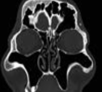
The first medical description of acromegaly was in 1886, by French neurologist Pierre Marie. This rare disease is usually caused by a pituitary adenoma, which has a typical phenotype of overgrowth, craniofacial disproportions and systemic complications, specially in the cardiovascular, respiratory and metabolic systems, secondary to excessive levels of growth hormone and insulin-like growth factor. Radiological detection (computed tomography and magnetic resonance imaging) of these craniofacial features is important, as they provide patients with an early diagnosis and treatment of disease.
Downloads
References
De Herder WW. The history of acromegaly. Neuroendocrinology. 2016;103:7-17.
Vilar L, Vilar CF, Lyra R, Lyra R, Naves LA. Acromegaly: Clinical features at diagnosis. Pituitary. 2017;20(1):22-32.
Chandna A, Islam N, Jabbar A, et al. Clinical features and outcome of surgery in 30 patients with acromegaly. J Pak Med Asoc. 2004;54(6):315-9.
Scacchi M, Cavagnini F. Acromegaly. Pituitary. 2006;9(4):297-303.
Karakıs D, Yılmaz B, Dogan A, Yetkin I, Bek B. The bite force and craniofacial morphology in patients with acromegaly : A pilot study. Med Oral Patol Oral Cir Bucal. 2014;19(1):e1-7.
Gosau M, Vogel C, Moralis A, Proff P, Kleinheinz J, Driemel O. Mandibular prognathism caused by acromegaly-a surgical orthodontic case. Head & Face Medicine. 2009;5:16.
Lavrentaki A, Paluzzi A, Wass JAH, Karavitaki N. Epidemiology of acromegaly: review of population studies. Pituitary. 2017; 20(1):4-9.
Buchfelder M, Schlaffer SM. The surgical treatment of acromegaly. Pituitary. 2017;20(1):76-83.
Ebner FH, Kürschner V, Dietz K, Bültmann E, Nägele T, Honegger J. Craniometric changes in patients with acromegaly from a surgical perspective. Neurosurg Focus. 2010;29(4):1-5.
Zada G, Cavallo L, et al. Transsphenoidal surgery in patients with acromegaly: Operative strategies for overcoming technically challenging anatomical variations. Neurosurg Focus. 2010;29:18-20.
Valenta LJ, Elias AN. Clinical acromegaly with undetectable growth hormone and hyperprolactinemia. J Natl Med Assoc. 1987;79(5):555-60.
Katznelson L, Atkinson JL, Cook DM, Ezzat SZ, Hamrahian AH, Miller KK, American Association of Clinical Endocrinologists. American Association of Clinical Endocrinologists medical guidelines for clinical practice for the diagnosis and treatment of acromegaly - 2011 update. Endocr Pract. 2011;17(Suppl 4):1-44.
Heireman S, Delaey C, Claerhout I, Decock CE. Restrictive extraocular myopathy: A presenting feature of acromegaly. Indian J Ophthalmol. 2011;59:517-9.
García JCF, Juan CS, Ballester AH. Aportación de la tomografía por emisión de positrones al diagnóstico de un caso de acromegalia. Endocrinol Nutr. 2008;55(4):175-7.
Lugo G, Pena L, Cordido F. Clinical manifestations and diagnosis of acromegaly. Int J Endocrinol. 2012;2012:540398.
Hodder SC, Rees JI, Oliver TB, Facey PE, Sugar AW. SPECT bone scintigraphy in the diagnosis and management
Downloads
Published
How to Cite
Issue
Section
License

This work is licensed under a Creative Commons Attribution-NonCommercial-ShareAlike 4.0 International License.
La Revista Colombiana de Radiología es de acceso abierto y todos sus artículos se encuentran libre y completamente disponibles en línea para todo público sin costo alguno.
Los derechos patrimoniales de autor de los textos y de las imágenes del artículo como han sido transferidos pertenecen a la Asociación Colombiana de Radiología (ACR). Por tanto para su reproducción es necesario solicitar permisos y se debe hacer referencia al artículo de la Revista Colombiana de Radiología en las presentaciones o artículos nuevos donde se incluyan.


