
Foreign Bodies of Atypical Presentation: Case Reports
DOI:
https://doi.org/10.53903/01212095.91Keywords:
Foreign bodies, Multidetector computed tomography, RadiologyAbstract
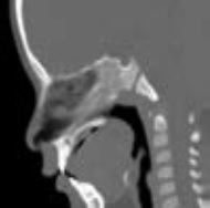
Objective: To show cases of foreign bodies of unconventional presentation diagnosed incidentally by various types of imaging studies in patients who did not refer and neither complained of a foreign body. Material and Methods: images from the archive of the radiology and diagnostic images service department were obtain from patients diagnosed of having a foreign body without a history related to this diagnosis. Results: Four clinical cases of patients of different ages are reported in whom foreign body was incidentally found in different anatomical locations and different imaging studies such as radiography and computed tomography. Conclusions: The nature of foreign bodies is diverse, being mainly of organic origin (dry wood). The clinical presentation will depend on the size, location and composition of the foreign body. The diagnosis can present several issues because they can present radiographic densities similar to the surrounding tissues. Computed tomography is the study of choice presenting limitations in the detection of foreign bodies of organic origin. In some cases only surgical exploration allows diagnosis and treatment.
Downloads
References
Javadrashid R, Fouladi DF, Golamian M, et al. Visibility of different foreign bodies in the maxillofacial region using plain radiography, CT, MRI and ultrasonography: an in vitro study. Dentomaxillofacial Radiol. 2015;44 (4):201402292.
Arango L, León L, Martínez D, Jurado G. Cuerpo extraño incidental en tracto gastrointestinal: Reporte de tres casos y revisión de la literatura. Rev Col Gastroenterol. 2011;26(4):316-27.
Kaviani F, Rashid R, Shahmoradi Z, Gholamian M. Detection of foreign bodies by spiral computed tomography and cone beam computed tomography in maxilofacial regions. J Dent Res Dent Clin Dent Prospects. 2014;8(3):166-71.
Sequiera Joyce B. H. Sripathi R, Mampilly MO, et al. Foreign body granuloma. J. Maxillofac. Oral Surg. 2014;13(3):351-4.
Lara C, Faba G, Caro J. Diagnóstico, manejo y actualización en cuerpo extraño aerodigestivo. Rev. Otorrinolaringol. Cir. Cabeza Cuello. 2008;68:309-18.
Jarraya M, Hayashi D, Villiers R, et al. Multimodality imaging of foreign bodies of the musculoskeletal system. AJR. 2014;203:W92-W102.
Peterson JJ, Bancroft LW, Kransdorf MJ. Wooden foreign bodies: imaging appearance. AJR Am J Roentgenol. 2002;178(3):557-62.
Horton LK, Jacobson JA, Powell A, et al. Sonography and radiography of softtissue foreign bodies. AJR. 2001;175:1155-9.
Esquivel DL, González GE. Diagnóstico diferencial por cuerpo extraño o patología fantasma en paladar duro en lactantes. Revisión de literatura y reporte de caso. Acta Odontol Colomb. 2017;7(2):85-93.
Kamble V, Mitra K, Dhote S. Un hallazgo radiográfico incidental: Torus palatinus. J Mahatma Gandhi Inst Med Sci. 2016;21:160-1.
Morales CH, Gómez F. Cuerpo extraño en el pericardio: extracción mediante videotoracoscopia. Rev. Colomb. Cir. 1997;12(4):264-6.
Soren S, Chaudhury S, Bakhla AK. Multiple self- inserted pins and nails in pericardium in a patient of schizophrenia: Case report and review. Ind Psychiatry J. 2015;24(1):82-7. 13. Supomo Darmawan H. An unusual foreign body in the heart: a case report. Ann Thorac Cardiovasc Surg. 2018;24(4):205-7.
Downloads
Published
How to Cite
Issue
Section
License

This work is licensed under a Creative Commons Attribution-NonCommercial-ShareAlike 4.0 International License.
La Revista Colombiana de Radiología es de acceso abierto y todos sus artículos se encuentran libre y completamente disponibles en línea para todo público sin costo alguno.
Los derechos patrimoniales de autor de los textos y de las imágenes del artículo como han sido transferidos pertenecen a la Asociación Colombiana de Radiología (ACR). Por tanto para su reproducción es necesario solicitar permisos y se debe hacer referencia al artículo de la Revista Colombiana de Radiología en las presentaciones o artículos nuevos donde se incluyan.


